Unveiling the Hidden Beauty of Adenomatoid Odontogenic Tumour Using CBCT: A Case Study

Dr. Navendra Jha1*, Dr. Fatima Injela Khan2, Dr. Faiza Khan MDS3, Dr. Sadiq Siddiqui MDS3

¹Assistant Professor, Department of Oral Medicine & Radiology, Seema Dental College and Hospital, Rishikesh, Uttarakhand
²Post Graduate Student, Department of Oral Medicine & Radiology, Institute of Dental Sciences, Bareilly, UP
³S.S Dental Clinic, Garur, Distt- Bageshwar, Uttarakhand

*Corresponding author

*Dr. Navendra Jha, Assistant Professor, Department of Oral Medicine & Radiology, Seema Dental College and Hospital, Rishikesh, Uttarakhand.

Abstract

In the maxillary, mandibular, and oral mucosa, odontogenic tumors (OT) are unique lesions that originate from mesenchymal and/or ectomesenchymal cells that are or were involved in the odontogenesis process.

Adenomatoid Odontogenic Tumour (AOT) refers to a benign tumour produced by the growth of epithelial remnants involved in the process of odontogenesis. Slow, expanded growth linked to certain included teeth, usually maxillary canines, is its defining feature.

It makes up 3% of all tumors that are odontogenic. These tumors' most distinctive characteristics are their location in the anterior maxilla and the appearance of internal calcifications on radiographs. It has a female predilection.

In this case study, we describe a 20-year-old female patient's radiographic characteristics, specifically using Cone Beam Computed Tomography. As of right now, very few case studies that use CBCT as an imaging modality and concentrate on radiographic aspects have been referenced in the literature.

In dentistry, CBCT is a leading-edge imaging technique that helps with precise treatment planning by offering a three-dimensional evaluation of odontogenic tumors. Because of its favorable prognosis and low recurrence rate, conservative surgery—either by enucleation or curettage—is the preferred course of treatment.

Keywords: Odontogenic tumour, Adenomatoid Odontogenic Tumour, Cone Beam Computed Tomography

Introduction

In the Indian population, adenomatoid odontogenic tumors stand fourth in its frequency. It is the most prevalent odontogenic tumor linked to teeth that have impacted.1 Given that it is most frequently linked to an impacted maxillary canine, the diagnosis is significant.2 Adenomatoid odontogenic tumors (AOTs) are epithelial tumors that exhibit duct-like features and whorled masses of cells amid a sparse fibrous stroma.3

Philipsen and Birn proposed the term "adenomatoid odontogenic tumor" in 1969. The World Health Organization (WHO) first used the term "adenomatoid odontogenic tumor" in 1971.4

AOT is more prevalent in the anterior side of the jaw than in the posterior region, and it is more common in the maxilla (64.3%) than in the mandible.5 Its distinguishing features can be accurately and elegantly evaluated with the aid of CBCT examination.6

The primary focus of this case study of a female patient aged 20 is the diagnostic characteristics of this tumor employing Cone Beam Computed Tomography as a three-plane radiographic imaging modality.

Case Report

A 20 year old female patient was referred for diagnostic imaging using CBCT as imaging modality for diagnosis. A Carestream Select 9300 CBCT machine (Carestream Dental LLC, Atlanta, GA, USA) was used to obtain her CBCT assessment. The patient was scanned using one of the imaging protocols that are compatible with this CBCT equipment and are listed below: 90 kVp, 8 mA, and 11–12 sec. With an 8x8 cm field of view, a 0.2 mm voxel size, and the patient's anatomy, the gadget determined the milliamp setting. The photos were evaluated using the Kodak Dental CS 3D Imaging Software V3.5.7.0 (Carestream Health Inc., St. Rochester, NY, USA). Visualizing these pictures in axial, sagittal, and coronal portions is known as multiplanar reconstruction.

While evaluating the 3-D image there was bone destruction noted extending from the 11 to the 15 region with impacted tooth 13 at right maxilla. (Figure 1)

Figure 1: Three- dimensional image of Right Maxilla

To determine the extent of the lesion, the precise dimensions of the tumor were measured using the measurement tool. It measured approximately 31.0 mm × 28.1 mm in its greatest dimensions. (Figure 2)

Figure 2: Panoramic image depicting the dimensions of the lesion.

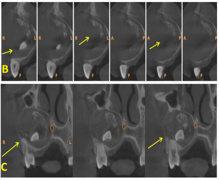
Figure 3: (A) Axial images (B)Cross sectional images (C) Coronal section

In the axial, coronal, and cross sections, we can observe a single, clearly defined unilocular radiolucency at the right anterior maxilla that extends from teeth 11 to 15 and involves the impacted tooth 52.

The pathology's internal structure revealed that it comprised several radiopacities or tiny calcifications that resembled snowflakes. The bone's labial cortical plate was perforated, and the palatal cortical plate was thinning. The two cortical plates were expanding. At the root apices of teeth 11, retained deciduous 52, 13, 14, and 15, root resorption was visible.There was displacement of the tooth 13. The pathology is involving the Right Nasal floor and Right Maxillary sinus. It was also involving the Nasopalatine nerve canal. In the dental findings a retained deciduous tooth 52 was present.

Discussion

Adenomatoid odontogenic tumors are regarded as benign, hamartomatous epithelial lesions of odontogenic origin as opposed to neoplasia.7 It has been proposed that this disease develops from remains of the dental lamina, despite evidence indicating tumor cells originate from the enamel's epithelium.3 Adenomatoid odontogenic tumors usually occur in females and young people.8 This lesion, which is connected to non-erupted canines, usually begins at the anterior region of the upper and lower jaw. Compared to patients who are male, female patients are twice as afflicted.6

There are three clinical variations of adenomatoid odontogenic tumors: follicular, extra-follicular, and peripheral.1 The majority of these tumors exhibit a variety of inductive alterations in the connective tissue, including eosinophilic and tiny calcification regions.9 Philipsen states that the AOT extra-follicular type is present in four distinct places: the middle third of the root, intra-radicular, periapical, and intra-osseous and unrelated to retained teeth.3

The gubernaculum dentis, also known as accessory dental lamina, is a preformed canal that extends from the dental follicle to the gum and permits the eruption of the permanent tooth during the mixed dentition period.2 It has been demonstrated that AOT originates from this canal and has an inductive effect on connective tissue.10 Additionally, some tumors may migrate along the gubernaculum dentis during tooth eruption, and it has been proposed that AOT may develop from epithelial debris at the crown of a permanent tooth.4

These lesions resemble other lesions of odontogenic origin on radiography. However, the type of tumor determines these features.5 They manifest as a distinct, bounded region that is connected to the crown or root of an impacted tooth in the follicular form.11

When a tooth erupts, the detected lesion is situated between or above its roots in the extra-follicular form.8 The peripheral form is characterized by tooth displacement in certain situations without resulting in root resorption, as well as thinning or resorption of the cerebral cortex. 10

Calcification exhibits as globular, spheroidal, leisegang rings, and irregular masses.11 They react well to a conservative treatment that consists of enucleation and curettage because of their non-invasive nature, sluggish growth, and capsule-like structure. There has been no evidence of recurrence.12

Conclusion

The facts of this instance were consistent with those of all the AOT report that has been citedto date. In order to properly grasp the tumor's features and aid in treatment planning, appropriate radiographic features are shown utilizing the soon-to-be popular CBCT as radiographic imaging.

References

  1. Virupakshappa D, Rajashekhara BS, Manjunatha BS, Das N (2014). Adenomatoid odontogenic tumour in a 20-year-old woman. BMJ Case Rep; 3(1):1- 4.
  2. Mutalik VS, Shreshtha A, Mutalik SS, Radhakrishnan R (2016). Adenomatoid odontogenic tumor: A unique report with histological diversity. J Oral Maxillofac Pathol; 16(1):118-21.
  3. Mohamed A, Singh AS, Raubenheimer EJ (2010). Adenomatoid odontogenic tumour: review of the literature and an analysis of 33 cases from South Africa. Int J Oral Maxillofac Surg; 39(9):843–6.
  4. Handschel JG, Depprich RA, Zimmermann AC, Braunstein S, Kübler NR (2005). Adenomatoid odontogenic tumor of the mandible: Review of the literature and report of a rare case. Head Face Med; 24(1):1-3.
  5. Garg D, Palaskar S, Shetty VP, Bhushan A (2009). Adenomatoid odontogenic tumor - hamartoma or true neoplasm: A case report. J Oral Sci; 51(1):155-9.
  6. Ide F (2010). Inter-radicular adenomatoid odontogenic tumor of the anterior mandible. J Oral Maxillofac Surg; 68(1):490–1.
  7. Motamedi MH, Shafeie HA, Azizi T (2005). Salvage of an impacted canine associated with an adenomatoid odontogenic tumour: a case report. Brit Dent J; 199(2):89–90.
  8. Komal K, Vibhakar A (2011). Mural adenomatoid odontogenic tumor in the mandible: A rare case. Int J Oral Maxillofac Pathol. 4(2):35–44.
  9. Acharya S, Goyal A, Rattan V, Vaiphei K, Kaur Bhatia S (2014). Dentigerous cyst or adenomatoid odontogenic tumor: clinical radiological and histopathological dilemma. Case Reports in Medicine. 2 (1):514-7.
  10. Shetty K, Vastardis S, Giannini P (2005). Management of an unusually large adenomatoid odontogenic tumor. Oral Oncology Extra. 41(2):316–18.
  11. Jham BC; Passos JB; Carmo MAVd (2006). Adenomatoid odontogenic tumor originated in the periodontal ligament. Oral Oncology Extra. 42(1):268–71.
  12. Shivali V; Pandey A; Khanna VD; Khanna P; Singh A., (2013). A rare case of extrafollicular adenomatoid odontogenic tumor in the posterior region of the mandible: misdiagnosed as residual cyst. Journal of International Oral Health. 5(5), 124–8.
TOP