
Simple Liver Cyst as A Differential Diagnosis of Right Adrenal Cyst
Murube Algarra, Carmen1*; Campos Martínez, Francisco Javier2; Calvo Duran, Antonio Enrique3; Falckenheiner Soria, Joshua Ernesto4; Vega Ruiz, Vicente5
¹Resident in general and digestive surgery at the University Hospital of Puerto Real (Cádiz, Spain). PhD student at the University of Cadiz.
²MD PhD. FEA in general and digestive surgery at the University Hospital of Puerto Real (Cádiz, Spain).
³MD PhD. Head of the General Surgery and Digestive System Section at the Puerto Real University Hospital (Cádiz, Spain)
⁴MD PhD .FEA of General Surgery and Digestive System at the Puerto Real University Hospital (Cádiz, Spain)
⁵MD PhD .Head of the General Surgery and Digestive System Unit at the Puerto Real University Hospital (Cádiz, Spain).
*Corresponding author
*Murube Algarra, Carmen, Resident in general and digestive surgery at the University Hospital of Puerto Real (Cádiz, Spain). PhD student at the University of Cadiz.
DOI: 10.55920/JCRMHS.2025.09.001400
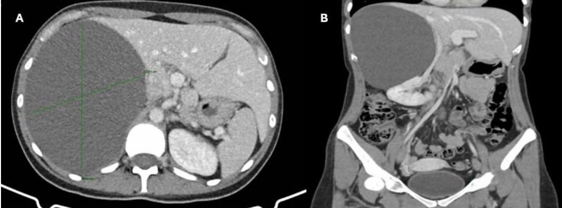
Figure 1: Abdominal CT scan showing a well-defined lesion in the right hepatic lobe, measuring 17x15.7x12cm (LxAPxT) (A) that displaces the right kidney medially and inferiorly (B).
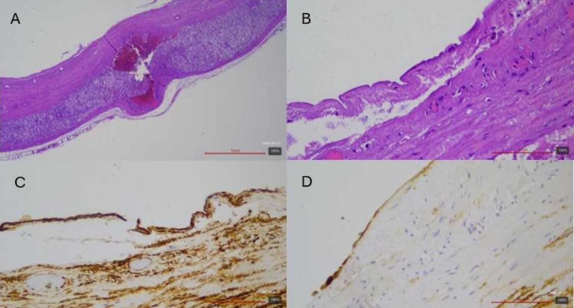
Figure 2: Histological sections of adrenal parenchyma showing a cystic cavity lined by flat endothelium expressing CD34 and D2-40, confirming its endothelial origin. A: 5x hematoxylin-eosin stain. B: 40x hematoxylin-eosin stain. C: CD34 immunohistochemistry. D: D2-40 immunohistochemistry
Image 1: Abdominal CT scan showing a well-defined lesion in the right hepatic lobe, measuring 17x15.7x12cm (LxAPxT) (A) that displaces the right kidney medially and inferiorly (B).
After ruling out infection by Echinococcus spp., the case was presented to the Hepatobiliary-pancreatic Tumors Committee. Due to the magnitude of the lesion and the associated secondary symptoms, surgical treatment was decided for possible symptomatic giant HSQ.
An exploratory laparoscopy was performed, revealing a cystic lesion located in the posterior lateral segments of the liver (segments VI-VII), with an approximate dimension of 15 x 15 cm. The lesion causes displacement of the inferior vena cava, the hepatic flexure of the colon, the kidney and right adrenal gland, as well as the right hepatic lobe. After complete dissection of the lesion, complete dependence on the adrenal gland without origin at the hepatic level is evident.
Given the findings, complete excision of the cyst was carried out by means of enucleation associated with a total adrenalectomy, due to the close relationship and fusion with the right adrenal gland, in relation to the probable adrenal origin of the lesion.
The postoperative period was without significant complications, and the patient was discharged on the third postoperative day.
The anatomopathological analysis confirmed the diagnosis of an endothelial type cyst of adrenal origin without histological characteristics of malignancy, with a positive immunohistochemical study for CD34 and D2-40 (Image 2).
Image 1: Histological sections of adrenal parenchyma showing a cystic cavity lined by flat endothelium expressing CD34 and D2-40, confirming its endothelial origin. A: 5x hematoxylin-eosin stain. B: 40x hematoxylin-eosin stain. C: CD34 immunohistochemistry. D: D2-40 immunohistochemistry