TNFAIP involved in PDGFB-PDGFRB signaling communication in the Chemotherapeutic response of Triple-negative Breast Cancer (TNBC)

Zhaoyang Qin1,#, Gangping Wang2,3,#,*

¹Department of General Surgery, The Rizhao People’s Hospital, 276800, Rizhao, China.
²Department of Pathology, the Fourth Affiliated Hospital of School of Medicine, & International School of Medicine, International Institutes of Medicine, Zhejiang University, 322000, Zhejiang, China.
³Central Laboratory, The Rizhao People’s Hospital, 276800, Rizhao, China.
#Equal contribution

*Corresponding author

*Gangping Wang, MD, Department of Pathology, the Fourth Affiliated Hospital of School of Medicine, and International School of Medicine, International Institutes of Medicine, Zhejiang University, Zhejiang, China.

Abstract

Background and Objective: Triple-negative breast cancer (TNBC) is a highly aggressive subtype known for its high recurrence rates, poor prognosis, lack of tumor predictive markers and potential therapeutic targets, and complex molecular mechanisms that have not yet been elucidated. TNFAIP has both carcinogenic and anticancer effects. This study highlights the key role of TNFAIP in PDGFB-PDGFRB signaling in TNBC chemotherapy and tumor angiogenesis by single-cell transcriptomic analysis.

Methods: We utilized the raw data from the GEO database hosted by NCBI to analyze single-cell transcriptomes. Additionally, we employed R software packages, such as Seurat (v.4.1.1), DoubletFinder (v.2.0.3), ClusterProfiler (v.4.2.2), CellChat (v.1.1.3), and other pertinent software, to perform a range of comprehensive analyses. These analyses encompassed quality control of single-cell transcriptome sequencing data, sample integration, dimensionality reduction, cell clustering and grouping, differential gene expression analysis, GO enrichment, and intercellular communication analysis.

Results: The results revealed a single-cell sequencing map of tumor lesion cell samples from TNBC before and after chemotherapy, identifying 8 cell subpopulations labeled as 8 primary cell types. Notably, the cell communication between endothelial cells and fibroblasts in TNBC stands out, surpassing other types of cell communication, suggesting a regulatory role between these two cell types. Through ligand-receptor communication probability analysis, it was determined that the PDGF signaling pathway between endothelial and fibroblasts in TNBC serves as a crucial ligand-receptor pathway. During chemotherapy in TNBC patients, the regulation of the PDGF signaling pathway might primarily be influenced by PDGFB-PDGFRB, which stimulates matrix proliferation, promotes tumor angiogenesis, and contributes to tumor metastasis. Furthermore, single-cell Pearson correlation analysis indicated a correlation between the TNFAIP and PDGF pathways, suggesting an association between TNFAIP and the endothelial cell-fibroblast PDGFB-PDGFRB signaling pathway.

Conclusions: Understanding the complex regulatory network involving TNFAIP in PDGFB-PDGFRB signaling, as well as its interaction with angiogenesis-related factors in TNBC chemotherapy response, offers valuable insights for developing new treatment strategies and identifying potential biomarkers for TNBC.

*Key Words: Triple-negative breast cancer, Sequence, Single-cell transcriptomes, PDGF, TNFAIP, Tumor angiogenesis, Bioinformatic analysis, Chemotherapy.

Introduction

Triple-negative breast cancer (TNBC) is a highly aggressive subtype known for its high recurrence rates, poor prognosis[1], lack of tumor predictive markers and potential therapeutic targets, and complex molecular mechanisms that have not yet been elucidated[2]. Platelet derived growth factor (PDGF) is essential for the development of the vasculature and the formation and remodeling of tumor blood vessels [3]. Recent Single-cell transcriptomic analysis also found that the PDGF signaling pathway serves as a possible therapeutic target for TNBC[4]. Platelet derived growth factor receptor beta (PDGFRB) and structural homologous proteins PDGFRA are members of the receptor tyrosine kinase (RTK) class III subfamily[5], which also includes receptors for SCF, M-CSF, and Flt-3 ligands[6]. Tumor angiogenesis plays a pivotal role in progression and metastasis of TNBC[7,8]. The hypoxic tumor microenvironment promotes angiogenesis through dysregulation of ncRNAs, including microRNAs and lncRNAs[9,10], and is involved in the tumor necrosis factor alpha-induced protein (TNFAIP) and NF-κB signaling pathways[11,12]. Tumor necrosis factor(TNF) is a cytokine with multiple functions, playing a crucial role in early inflammation, angiogenesis, immune regulation, immune defense, immune cell differentiation, cancer biology, and the prevention of malignant cell growth during the immune response[13]. TNFAIP is a protein associated with inflammation and tumors and has been found to be involved in the occurrence and development of various types of tumors. TNFAIP1 has both carcinogenic and anticancer effects. Research has shown that low-oxygen conditions can stimulate changes in tumor cell TNFAIP, which in turn regulates angiogenesis through multiple mechanisms, including promoting endothelial cell migration and proliferation, regulating angiogenesis-related signaling pathways, regulating endothelial cell adhesion and permeability, and being associated with tumor invasion and prognosis. TNFAIP has been found to regulate the angiogenesis of TNBC cells by interacting with angiogenesis-related factors such as MMP and VEGF. In addition, TNFAIP can promote angiogenesis by activating inflammatory responses and increasing endothelial cell permeability. Angiogenesis is one of the important factors for the growth and metastasis of TNBC. At present, the research on the TNFAIP signaling pathway in TNBC is relatively limited, and further research on its signaling pathway is still needed. This study investigated the regulatory mechanism of TNFAIP in PDGFB-PDGFRB signaling communication in the chemotherapy response of TNBC.

Materials and Methods

Single cell transcriptome data acquisition and analysis process

The single-cell transcriptome sequencing data used in this study are all publicly available raw data included in the GEO database under NCBI, with the inclusion number GSE263995. The download link is https://www.ncbi.nlm.nih.gov/geo/query/acc.cgi?acc=GSE263995[14]. Download the single cell transcriptome sequencing data of tumor focus cell samples of TNBC patients before and after chemotherapy from the GEO database, and use R packages such as Seurat (v.4.1.1), DoubletFinder (v.2.0.3), ClusterProfiler (v.4.2.2), CellChat (v.1.1.3) and other relevant software in the R software package to conduct a series of in-depth analysis, including quality control, sample integration, dimension reduction analysis, cell grouping, differential gene analysis, GO enrichment and intercellular communication analysis.

Quality control of single-cell transcriptome sequencing data

In order to control the quality of single-cell transcriptome sequencing data, a series of quality filters were performed on the raw data. According to the recommendations of the Seurat software package, select the threshold for filtering parameters to remove cells with too few expressed genes and cells containing too many unique molecular identifiers (UMIs). Simultaneously use the R software package DoubletFinder to predict double cells. Through the above methods, low-quality cells and double cells are filtered, while high-quality single cells that meet quality control standards are retained and used for subsequent analysis.

Integration and standardization analysis of single cell transcriptome sequencing data

Using the R software package Seurat for data integration analysis of different samples in single-cell transcriptome sequencing data, merge all Seurat objects constructed based on retained UMIs gene expression matrices into one Seurat object. Then, using the SCTransform (v.0.3.4) algorithm in the R software package Seurat, the single-cell transcriptome sequencing data was standardized, normalized, and feature analyzed based on regularized negative binomial regression.

Cell clustering and grouping annotations

Using the principal component analysis (PCA) algorithm in the R software package Seurat, linear dimensionality reduction was performed on the top 3000 variable genes. According to the built-in standard steps, select 1-50 highly variable principal components (PCs) and use the uniform manifold approximation and projection (UMAP) method to perform unsupervised clustering and clustering of cells. Using the software package SingleR (v.2.2.0) to combine the gene expression of these cell populations with CellMarker 2.0[15](http://bio-bigdata.hrbmu.edu.cn/CellMarker or http://117.50.127.228/CellMarker/) PanglaoDB[16]( https://panglaodb.se/index.html )Wait for database analysis to define cell types. Use the FindAllMarkers feature in the Seurat software package to validate the marker genes of each cell subpopulation and perform cell annotation.

Differential analysis and GO enrichment analysis

Further enrichment analysis of GO (Gene Ontology) biological processes (BP) was performed using R software package ClusterProfiler on cell subpopulation specific high expression genes identified by FindAllMarkers or FindMarkers functionality in Seurat.

Analysis of intercellular communication

Using the R software package CellChat (v1.1.0), follow the workflow provided by GitHub (https://github.com/sqjin/CellChat/blob/master/tutorial/CellChat-vignette.Rmd)[17] Analyze cell-cell communication based on the expression levels of ligands and receptors in different cell types. In short, load the standardized single-cell data into CellChatDB software and perform preprocessing functions such as "identifiyOverExpressedGenes", "identifiyOverExpressedInteractions", and "projectData". We chose the "secretion signaling" related pathway and used the "ligand receptor interaction" recorded in the database as the cell communication information for prediction. In formal analysis, standard parameters and fixed random seeds are used to calculate the core functions computeCommonProb, computeCommonProbPathway, and aggregateNet. Finally, in order to determine the sender (expressed ligand) and receiver (expressed receptor) in cellular communication, the function "netAnalysis_SignalingRole" was applied in the "netP" data for calculation.

Statistical Methods

Statistical analysis was conducted using R software (v4.2.1), and t-test was used for differential analysis. | logFC |>1 is the threshold for expression differences, where | logFC |>1 is an upregulated gene, | logFC |<-1 is a downregulated gene, and P<0.05 is statistically significant.

Results

Cell Grouping and Identification

We used the R software package DoubletFinder to predict and filter double cells from the raw single-cell transcriptome sequencing data of tumor lesion cell samples from TNBC patients before and after chemotherapy, which were included in the GEO database under the NCBI (GSE263995, https://www.ncbi.nlm.nih.gov/geo/query/acc.cgi?acc=GSE263995). High-quality single cells that met quality control standards were retained for subsequent analysis. Using the R software package Seurat to perform data integration analysis of different samples in single-cell transcriptome sequencing data, based on the SCTransform (v.0.3.4) algorithm, regularized negative binomial regression was used to standardize, normalize, and analyze the characteristics of single-cell transcriptome sequencing data; The PCA algorithm in the R software package Seurat was used to perform linear dimensionality reduction on the top 3000 variable genes, and 1-50 highly variable principal components (PCs) were selected. The UMAP method was used to perform unsupervised clustering and grouping of cells. CellMarker 2.0 was used to annotate the cell clusters, resulting in the identification of eight cell subpopulations (Figure 1A). Based on the characteristic gene expression levels of cell types, eight cell subpopulations were labeled as eight major cell types, including epithelial cells, endothelial cells, fibroblasts, T cells, B cells, macrophages, smooth muscle cells, and chondrocytes (Figure 1B). These cells constitute the single-cell atlas of tumor lesion cell samples from triple-negative breast cancer patients before and after chemotherapy. Next, for the genes specifically highly expressed in these cell subsets identified by the FindAllMarkers or FindMarkers functions in the R software package Seurat, we further used the R software package ClusterProfiler to perform GO enrichment analysis on the characteristic expression genes of cell types. The dimensionality reduction clustering and grouping of single-cell transcriptome data resulted in the following key biological processes, including T cell receptor signaling pathway, small GTPase-mediated signal transduction, regulation of T cell activation, regulation of biological stimulus response, phagocytic disease, muscle system process, muscle contraction, lymphocyte-mediated immunity, keratinocyte differentiation, immune response regulatory signaling pathway, extracellular structure organization, extracellular matrix organization, external encapsulation structure organization, epidermal development, epidermal cell differentiation, endothelial development, cell matrix adhesion, bone mineralization, biological mineral organization development, B cell receptor signaling pathway, B cell proliferation, B cell activation, etc. (Figure 1C). These processes are basically consistent with the functions of each cell cluster, proving that the grouping annotation is good.

Figure 1: Dimensionality reduction clustering and clustering of TNBC single-cell transcriptome data. (A) Expression levels of characteristic genes based on cell types.(B) Single cell sequencing map of tumor lesion cell samples before and after chemotherapy, with 8 cell subgroups labeled as 8 main cell types. (C) GO enrichment analysis of characteristic expressed genes in different cell types, dimensionality reduction clustering and clustering of single-cell transcriptome data, to identify key biological processes of signaling pathways.

Communication between endothelial cells and fibroblasts plays a regulatory role in TNBC

To further explore the interactions between endothelial cell-fibroblast populations in tumor lesion cell samples before and after chemotherapy in TNBC patients, we extracted cell populations from single-cell sequencing data maps, used the R package CellChat to analyze cell-cell communication networks, loaded standardized single-cell data into the CellChatDB software, and performed preprocessing functions such as "identifyOverExpressedGenes", "identifyOverExpressedInteractions", and "projectData" calculations. We used standard parameters and fixed random seeds to perform core functions such as computeCommunProb, computeCommunProbPathway, and aggregateNet calculations. We applied the function "netAnalysis_signalingRole" in the "netP" data for calculation. The results of the number and intensity of interactions between signaling output and receiving cell populations are shown in Figure 2A. The results show that the communication between endothelial cells and fibroblasts is the most significant, significantly higher than other cell-cell communication, suggesting that the communication between endothelial cells and fibroblasts plays an important role before and after chemotherapy. In the cell proportion diagram, endothelial cells and fibroblasts showed a significant increase and decrease trend before and after chemotherapy, indicating that there may be a regulatory process of intercellular communication between endothelial cells and fibroblasts (Figure 2B).

Figure 2: Communication network interactions between cell populations shows that communication between endothelial cells and fibroblasts plays a regulatory role in TNBC chemotherapy. (A) Number and intensity of intercellular interactions. (B) Change of cell proportion of tumor focus cell samples in TNBC patients pre and post chemotherapy.

PDGF signaling pathway is a key ligand receptor pathway of endothelial fibroblasts in TNBC by ligand receptor communication probability analysis

By calculating the communication probability at the ligand-receptor level and the communication probability at the signal pathway level, we obtained the aggregated communication network between cells (Figure 3A). The analysis of the ligand-receptor communication probability between endothelial cells and fibroblasts revealed that the PDGF signaling pathway is a key pathway for the ligand-receptor communication between endothelial cells and fibroblasts in TNBC (Figure 3B). PDGF is a paracrine growth factor that activates signaling pathways through binding to its receptors, participating in processes such as cell proliferation, migration, survival, and vascular regulation. PDGF receptors include two types, PDGFRA and PDGFRB, which correspond to the binding of PDGFA, PDGFB, and PDGFAB, respectively. When PDGF binds to its receptor, it causes the receptor to dimerize and activates the activity of receptor tyrosine kinase through phosphorylation. Abnormal activation or mutation of the PDGF signaling pathway is involved in the onset and progression of various diseases, including carcinogenesis, pulmonary fibrosis, and atherosclerosis. Activated PDGF receptors regulate multiple downstream signaling pathways, including PI3K/Akt, Ras/Erk, JAK/STAT, and Src, through tyrosine phosphorylation. Current research shows that it can stimulate interstitial proliferation and promote tumor angiogenesis, and is involved in the process of tumor metastasis. Endothelial cells, vascular smooth muscle cells, activated monocytes, and macrophages can all produce PDGF. PDGF is a growth stimulator for mesenchymal cells such as fibroblasts and smooth muscle cells, and can also induce vascular proliferation in vivo by activating macrophages to synthesize bFGF and TGF-β pathways, which then act on endothelial cells. During this process, it may promote the growth and migration of fibroblasts, and the regulation of its expression level has great potential in the treatment of TNBC. A deep understanding of the PDGF signaling pathway aids in revealing its pathological mechanisms of related diseases and provide new targets and strategies for disease treatment.
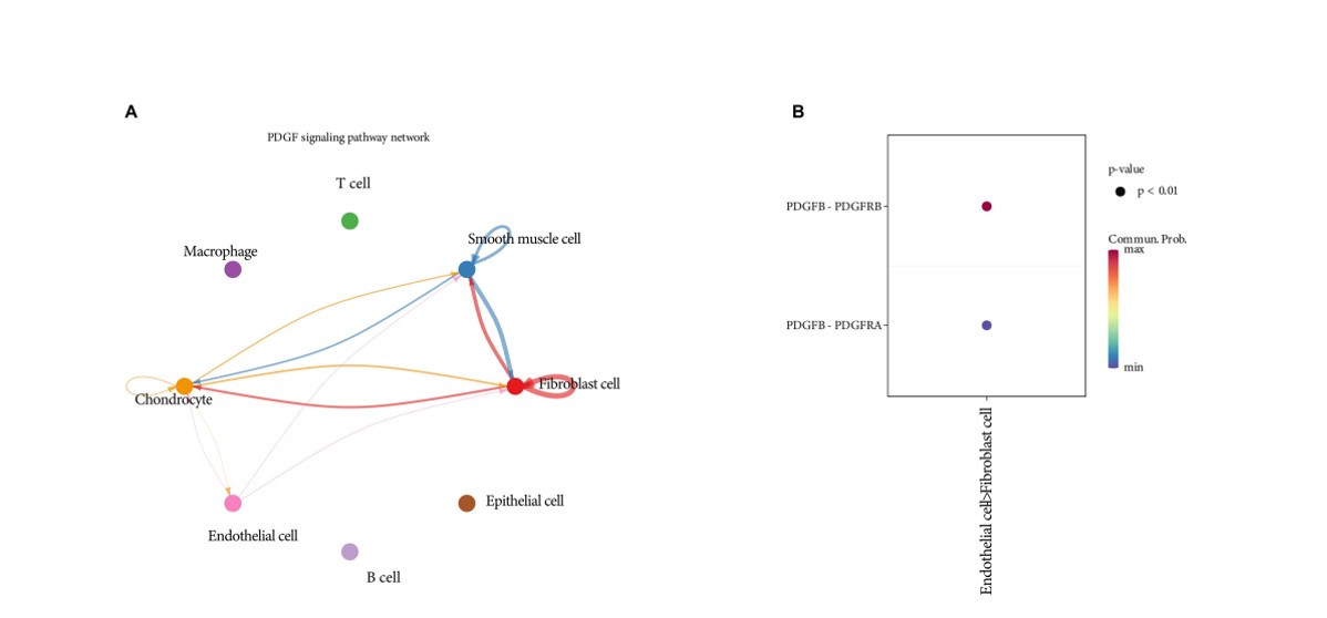
Figure 3: Probability of ligand receptor pair communication between endothelial cells and fibroblasts shows that the PDGF signaling pathway is a key ligand receptor pathway of endothelial fibroblasts in TNBC. (A) The aggregated communication network between cells by calculating the communication probability at the ligand receptor level and the communication probability at the signaling pathway level. (B) PDGF signaling pathway is a key ligand receptor pathway of endothelial fibroblasts.

The regulation of the PDGF signaling pathway may primarily be governed by the PDGFB-PDGFRB ligand-receptor pair during TNBC patients chemotherapy

We conducted an analysis of the changes in the PDGF signaling pathway in TNBC patients before and after chemotherapy, encompassing both intercellular communication within the PDGF signaling pathway and the contribution analysis of PDGF signaling pathway ligand-receptor pairs. The results indicate that during chemotherapy for triple-negative breast cancer, the PDGF signaling pathway extensively interacts across various cell populations, including endothelial cells, fibroblasts, smooth muscle cells, and chondrocytes(Figure 4A). This suggests that PDGF signaling targets a wide range of cells and exerts broad effects. Notably, the contribution of PDGFB-PDGFRB ligand-receptor pairs is notably stronger compared to other ligand-receptor pairs(Figure 4B). Therefore, the regulation of the PDGF signaling pathway may primarily be governed by the PDGFB-PDGFRB ligand-receptor pair.

Figure 4: Changes of PDGF signaling pathway pre or post chemotherapy in TNBC patients. (A) The PDGF signaling pathway network during the healing process. (B) Contribution of PDGF signaling pathway ligand receptor pairs.

TNFAIP correlated with PDGFB-PDGFRB signaling pathway of endothelial fibroblas in TNBC chemotherapy

TNFAIP is a protein linked to inflammation and tumors, having been found to associated with the occurrence and progression of various tumor types, including TNBC. Angiogenesis stands as a crucial factor in the growth and metastasis of TNBC. Studies reveal that hypoxic conditions can stimulate tumor cells to produce and release TNFAIP, subsequently modulating angiogenesis via multiple mechanisms. These include enhancing the migration and proliferation of endothelial cells, regulating signaling pathways related to angiogenesis, and modulating the adhesion and permeability of endothelial cells. Furthermore, TNFAIP can facilitate angiogenesis by triggering inflammatory responses and elevating endothelial cell permeability. We performed a visual analysis of gene expression within the TNFAIP family. Post-chemotherapy, the expression level of TNFAIP increases(Figure 5A) and is highly expressed in fibroblasts(Figure 5B), indicating that the TNFAIP gene may be involved in the tumor chemotherapy process of TNBC patients, regulating fibroblast proliferation and migration to exert its effect. To delve deeper into the communication between TNFAIP and endothelial-fibroblast interactions, we conducted a single-cell two-gene Pearson correlation analysis focusing on the PDGFB-PDGFRB receptor pair, a pivotal pathway in PDGF communication. The results yielded a p-value of 4.5e-8 (p<0.001), indicating a significant correlation between TNFAIP and the PDGF signaling pathway. Interfering with the PDGFB-PDGFRB receptor pair in the TNFAIP and endothelial-fibroblast PDGF signaling pathway holds therapeutic potential.

Figure 5: TNFAIP correlated with PDGFB-PDGFRB signaling pathway of endothelial fibroblas in TNBC chemotherapy. (A) Elevated expression level of TNFAIP after TNBC chemotherapy. (B) TNFAIP has a communication relationship with key receptors involved in PDGFB-PDGFRB communication in the endothelial fibroblast PDGF pathway.

Discussion

Triple-negative breast cancer (TNBC) exhibited negative expression of estrogen receptor (ER), progesterone receptor (PR), and human epidermal growth factor receptor 2 (HER2), and is highly heterogeneous, with complex molecular mechanisms that have not yet been elucidated[18]. TNBC has an increased level of angiogenesis, which is antagonized by chemotherapy, and exhibits a high degree of aggressiveness and is associated with poor prognosis[19], early metastasis and limited treatment options[20].

Tumor angiogenesis and metastasis are the most cause of death in TNBC[21], and the PDGF complex formed by isoforms and receptors plays a significant role in regulating the growth and survival of various tumor cells[22]. PDGF signaling pathway is frequently dysregulated in cancer, and the expression of PDGF receptor is associated with adverse prognostic factors in TNBC[23]. Angiogenesis plays a pivotal role in tumor growth, metastasis, thus emerging as an appealing target for therapeutic intervention in TNBC[24]. An important participant in regulating angiogenesis in TNBC is TNFAIP, which mediates the signaling pathway of NF-κB signaling pathway activation [25]. The deregulation of this signaling pathway leads to abnormal angiogenesis, tumor growth, and metastasis[26] [27]. TNFAIP is an inducible protein of TNF-α, a cytokine associated with inflammation[13]. In TNBC, TNFAIP may promote the development of TNBC by activating inflammatory signaling pathways and altering tumor blood vessels[7] [28]. TNFAIP can be used as a downstream effector of TNF-α signaling to regulate the proliferation, migration, and invasion of tumor cells[29]. TNFAIP has been found to interact with angiogenic factors such as VEGF. TNFAIP can affect the angiogenesis ability of TNBC by regulating the expression and activity of these factors[30]. Our research results show that cell communication between endothelial cells and fibroblasts in TNBC is prominent, exceeding other types of intercellular communication, indicating that there is a regulatory effect between these two cell types. Through ligand-receptor communication probability analysis, we determined that the PDGF signaling pathway between endothelial cells and fibroblasts in TNBC is a key ligand-receptor pathway. During chemotherapy for TNBC patients, the regulation of the PDGF signaling pathway may be mainly influenced by PDGFB-PDGFRB, which stimulates matrix proliferation, promotes tumor angiogenesis, and facilitates tumor metastasis[23]. In addition, single-cell Pearson correlation analysis showed a correlation between TNFAIP and PDGF pathways, indicating a link between TNFAIP and the PDGFB-PDGFRB signaling pathway in endothelial cell fibroblasts. This expansion of the regulatory theme provides insights into the specific role of TNFAIP in participating in the endothelial cell-fibroblast cell communication PDGFB-PDGFRB regulation of TNBC vascular expression and activity. This has expanded the understanding of the complex regulatory network involving TNFAIP and PDGF in response to hypoxic conditions and their impact on tumor angiogenesis[31].

In recent years, the concept of vascular normalization has become a promising strategy in TNBC chemotherapy[28,32]. The vascular normalization strategy can improve tumor perfusion in TNBC, enhance drug delivery, overcome treatment resistance[28], and enhance therapeutic efficacy[25]. The hypoxic microenvironment of tumors regulates angiogenesis[33,34], and TNFAIP has emerged as a crucial player in the context. The TNFAIP signaling pathway is participated in intercellular communication among endothelial fibroblasts in TNBC. The regulation of PDGFB-PDGFRB is closely related to angiogenesis regulation, providing valuable insights for developing new treatment strategies and the identification of potential biomarkers for TNBC[35]. These findings may translate into clinical practice and improve the efficacy of standard chemotherapy for TNBC. It should be noted that these results are only preliminary findings and more experimental studies are needed to verify and further explore the exact signaling pathway of TNFAIP in TNBC. Future research will help to more fully reveal the mechanism of TNFAIP in TNBC and provide new targets for the development of TNFAIP treatment strategies.

Conclusion

The intercellular communication between endothelial cells and fibroblasts in TNBC before and after chemotherapy mainly occurs through the PDGFB-PDGFRB key ligand-receptor pathway of PDGF, and there is a correlation between TNFAIP and the endothelial cell-fibroblast PDGFB-PDGFRB signaling pathway. Understanding the complex interactions involved in the regulation of TNBC vascularization by the endothelial cell-fibroblast PDGFB-PDGFRB signaling pathway during chemotherapy provides valuable insights into the regulatory mechanisms of tumor angiogenesis in TNBC. Intervention for these molecular interactions is a promising approach that is expected to develop new treatment strategies and personalized treatment options for patients with TNBC, further research and translation into effective clinical interventions, and improve the prognosis for patients with TNBC.

Ethics statement:

The study was approved by the Institutional Ethics Committee of the Fourth Affiliated Hospital, Zhejiang University School of Medicine, China (approval no. K2022011), and the Institutional Ethics Committee of the Medical Ethics Committee of Rizhao People's Hospital, China (approval no. 2021-KY-42). The study scRNA-seq data and bulk RNA-seq profiles from TNBC samples obtained from the GEO database did not require the approval of an ethics committee or informed consent as these data are publicly available.

Acknowledgments:

We thank the People's Hospital of Rizhao City, Shandong Province for providing fund matching support for this study. We appreciate the valuable contributions of the research group, especially Dr. Huifang Shi, and Dr. Lei Chen. The results of enrichment analysis were visualized via the GEO database under the NCBI platform (GSE263995, https://www.ncbi.nlm.nih.gov/geo/query/acc.cgi?acc=GSE263995).

Author contributions:

All authors were actively involved in the work on this manuscript. GPW designed the study; ZYQ performed bioinformatic analyses and image quantification; ZYQ and GPW carried out data interpretation and manuscript writing. All authors reviewed the manuscript and final acceptance of the submission.Author contributions.

Financial Support:

This work was supported by the Shandong Provincial Natural Science Foundation (ZR2020MH319) .

Conflict of Interest:

The authors declare that they have no competing interest.

Data availability statement:

The datasets analyzed for this study can be found in the GEO database (GSE263995, https://www.ncbi.nlm.nih.gov/geo/query/acc.cgi?acc=GSE263995).

References

  1. Acevedo-Díaz A, Morales-Cabán BM, Zayas-Santiago A, Martínez-Montemayor MM, Suárez-Arroyo IJ. SCAMP3 Regulates EGFR and Promotes Proliferation and Migration of Triple-Negative Breast Cancer Cells through the Modulation of AKT, ERK, and STAT3 Signaling Pathways. Cancers (Basel). 2022;14:2807.
  2. Gm A, E T, P M, Ss M, Kfa S. The Anticancer Effects of Flavonoids through miRNAs Modulations in Triple-Negative Breast Cancer. Nutrients [Internet]. 2021 [cited 2023 Jun 14];13. Available from: https://pubmed.ncbi.nlm.nih.gov/33916931/
  3. Guérit E, Arts F, Dachy G, Boulouadnine B, Demoulin J-B. PDGF receptor mutations in human diseases. Cell Mol Life Sci. 2021;78:3867–81.
  4. Foutadakis S, Kordias D, Vatsellas G, Magklara A. Identification of New Chemoresistance-Associated Genes in Triple-Negative Breast Cancer by Single-Cell Transcriptomic Analysis. Int J Mol Sci. 2024;25:6853.
  5. Paolini C, Agarbati S, Benfaremo D, Mozzicafreddo M, Svegliati S, Moroncini G. PDGF/PDGFR: A Possible Molecular Target in Scleroderma Fibrosis. Int J Mol Sci. 2022;23:3904.
  6. Zou X, Tang X-Y, Qu Z-Y, Sun Z-W, Ji C-F, Li Y-J, et al. Targeting the PDGF/PDGFR signaling pathway for cancer therapy: A review. Int J Biol Macromol. 2022;202:539–57.
  7. Ayoub NM, Jaradat SK, Al-Shami KM, Alkhalifa AE. Targeting Angiogenesis in Breast Cancer: Current Evidence and Future Perspectives of Novel Anti-Angiogenic Approaches. Front Pharmacol. 2022;13:838133.
  8. Yadav K, Lim J, Choo J, Ow SGW, Wong A, Lee M, et al. Immunohistochemistry study of tumor vascular normalization and anti-angiogenic effects of sunitinib versus bevacizumab prior to dose-dense doxorubicin/cyclophosphamide chemotherapy in HER2-negative breast cancer. Breast Cancer Res Treat. 2022;192:131–42.
  9. Oncology and Translational Medicine [Internet]. [cited 2024 Nov 25]. Available from: https://journals.lww.com/otm/fulltext/9900/perspectives_on_the_potential_application_of.68.aspx
  10. Wu Y, Song H, Shi H, Wang G. Title: LncRNA regulates the progression of Triple-negative breast cancer.
  11. Deepak KGK, Vempati R, Nagaraju GP, Dasari VR, S N, Rao DN, et al. Tumor microenvironment: Challenges and opportunities in targeting metastasis of triple negative breast cancer. Pharmacol Res. 2020;153:104683.
  12. Naik A, Decock J. Lactate Metabolism and Immune Modulation in Breast Cancer: A Focused Review on Triple Negative Breast Tumors. Front Oncol. 2020;10:598626.
  13. Wang X, Yang C, Körner H, Ge C. Tumor Necrosis Factor: What Is in a Name? Cancers. 2022;14:5270.
  14. GEO Accession viewer [Internet]. [cited 2024 Nov 25]. Available from: https://www.ncbi.nlm.nih.gov/geo/query/acc.cgi?acc=GSE263995
  15. Hu C, Li T, Xu Y, Zhang X, Li F, Bai J, et al. CellMarker 2.0: an updated database of manually curated cell markers in human/mouse and web tools based on scRNA-seq data. Nucleic Acids Res. 2022;51:D870–6.
  16. Franzén O, Gan L-M, Björkegren JLM. PanglaoDB: a web server for exploration of mouse and human single-cell RNA sequencing data. Database (Oxford). 2019;2019:baz046.
  17. CellChat/tutorial/CellChat-vignette.Rmd at master · sqjin/CellChat [Internet]. GitHub. [cited 2024 Nov 25]. Available from: https://github.com/sqjin/CellChat/blob/master/tutorial/CellChat-vignette.Rmd
  18. Ferrari P, Scatena C, Ghilli M, Bargagna I, Lorenzini G, Nicolini A. Molecular Mechanisms, Biomarkers and Emerging Therapies for Chemotherapy Resistant TNBC. Int J Mol Sci. 2022;23:1665.
  19. Qu F, Wang G, Wen P, Liu X, Zeng X. Knowledge mapping of immunotherapy for breast cancer: A bibliometric analysis from 2013 to 2022. Hum Vaccin Immunother. 2024;20:2335728.
  20. Tsang JY, Tse GM. Update on triple-negative breast cancers - highlighting subtyping update and treatment implication. Histopathology. 2023;82:17–35.
  21. Ozer LY, Fayed HS, Ericsson J, Al Haj Zen A. Development of a cancer metastasis-on-chip assay for high throughput drug screening. Front Oncol. 2024;13:1269376.
  22. Kim S, You D, Jeong Y, Yoon SY, Kim SA, Lee JE. Inhibition of platelet-derived growth factor C and their receptors additionally increases doxorubicin effects in triple-negative breast cancer cells. Eur J Pharmacol. 2021;895:173868.
  23. Jansson S, Aaltonen K, Bendahl P-O, Falck A-K, Karlsson M, Pietras K, et al. The PDGF pathway in breast cancer is linked to tumour aggressiveness, triple-negative subtype and early recurrence. Breast Cancer Res Treat. 2018;169:231–41.
  24. Maj E, Papiernik D, Wietrzyk J. Antiangiogenic cancer treatment: The great discovery and greater complexity (Review). International Journal of Oncology. 2016;49:1773.
  25. Liu Z, Zhang H, Sun L, Zhu K, Lang W. miR-29c-3p Increases Cell Viability and Suppresses Apoptosis by Regulating the TNFAIP1/NF-κB Signaling Pathway via TNFAIP1 in Aβ-Treated Neuroblastoma Cells. Neurochem Res. 2020;45:2375–84.
  26. Frontiers | Vascular Normalization: A New Window Opened for Cancer Therapies [Internet]. [cited 2023 Jun 15]. Available from: https://www.frontiersin.org/articles/10.3389/fonc.2021.719836/full
  27. Ayoub NM, Jaradat SK, Al-Shami KM, Alkhalifa AE. Targeting Angiogenesis in Breast Cancer: Current Evidence and Future Perspectives of Novel Anti-Angiogenic Approaches. Frontiers in Pharmacology [Internet]. 2022 [cited 2023 Jul 5];13. Available from: https://www.ncbi.nlm.nih.gov/pmc/articles/PMC8913593/
  28. Wang Z, Li Z, Sun Z, Wang S, Ali Z, Zhu S, et al. Visualization nanozyme based on tumor microenvironment “unlocking” for intensive combination therapy of breast cancer. Sci Adv. 2020;6:eabc8733.
  29. F B. TNF-alpha in promotion and progression of cancer. Cancer metastasis reviews [Internet]. 2006 [cited 2024 May 16];25. Available from: https://pubmed.ncbi.nlm.nih.gov/16951987/
  30. G K, D K. Role of TNF/TNFR in autoimmunity: specific TNF receptor blockade may be advantageous to anti-TNF treatments. Cytokine & growth factor reviews [Internet]. 2002 [cited 2024 Aug 23];13. Available from: https://pubmed.ncbi.nlm.nih.gov/12220546/
  31. Hernández-Vargas H, Rodríguez-Pinilla SM, Julián-Tendero M, Sánchez-Rovira P, Cuevas C, Antón A, et al. Gene expression profiling of breast cancer cells in response to gemcitabine: NF-kappaB pathway activation as a potential mechanism of resistance. Breast Cancer Res Treat. 2007;102:157–72.
  32. Lahooti B, Poudel S, Mikelis CM, Mattheolabakis G. MiRNAs as Anti-Angiogenic Adjuvant Therapy in Cancer: Synopsis and Potential. Frontiers in Oncology [Internet]. 2021 [cited 2023 Jun 11];11. Available from: https://www.ncbi.nlm.nih.gov/pmc/articles/PMC8695604/
  33. Liu J, Ai X, Cabral H, Liu J, Huang Y, Mi P. Tumor hypoxia-activated combinatorial nanomedicine triggers systemic antitumor immunity to effectively eradicate advanced breast cancer. 2021;273:120847.
  34. Ma S, Zhao Y, Lee WC, Ong L-T, Lee PL, Jiang Z, et al. Hypoxia induces HIF1α-dependent epigenetic vulnerability in triple negative breast cancer to confer immune effector dysfunction and resistance to anti-PD-1 immunotherapy. Nat Commun. 2022;13:4118.
  35. Zhao Y, Yu X, Li J. Manipulation of immune‒vascular crosstalk: new strategies towards cancer treatment. Acta Pharm Sin B. 2020;10:2018–36.
TOP