Phagocytosis of Activated Platelets by Monocytes and Neutrophils in Peripheral Blood

Kayenne Tan Qi En1*, Gloria Chen Yuquan1, Dr Yap Eng Soo1,2, Dr Cheryl XQ Lim2

¹Department of Laboratory Medicine, Ng Teng Fong General Hospital, Singapore.

²Department of Laboratory Medicine, National University Hospital, Singapore.

*Corresponding author

*Kayenne Tan Qi En, Department of Laboratory Medicine, Ng Teng Fong General Hospital, Singapore

Clinical Image

A 73-year-old patient with a history of ischaemic heart disease, atrial fibrillation, dyslipidaemia and gout was admitted to the hospital for fever and shortness of breath. Prior to admission, the patient was on Apixaban, Atorvastatin and Allopurinol. Complete blood count done revealed leukocytosis with a total white blood cell (WBC) count of 18.9 x 10⁹/L. The differential white blood cell count is as follows: absolute neutrophil count 15.4 x 10⁹/L, absolute monocytes count 1.0 x 10⁹/L, absolute lymphocyte count 1.9 x 10⁹/L, absolute eosinophil count 0.5 x 10⁹/L and absolute basophil count 0.1 x 10⁹/L. Haemoglobin and platelet count were within normal ranges and were 14.3g/dL and 177 x 109/L respectively.

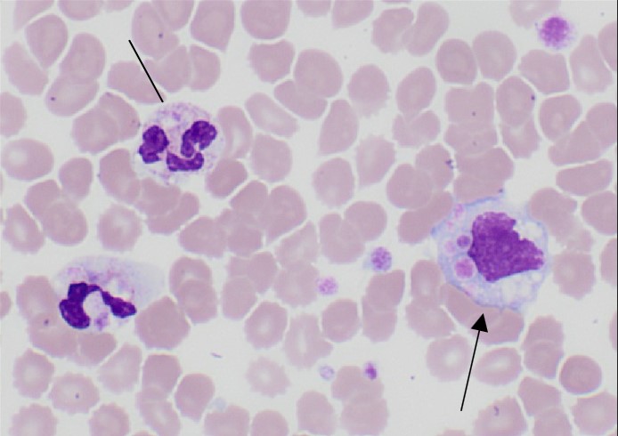
The patient’s blood count was flagged up due to leukocytosis and a peripheral blood film (PBF) was done. His PBF confirmed leukocytosis with neutrophilia with neutrophils displaying toxic vacuolations. The most striking feature was that of inclusion bodies within the cytoplasm of neutrophils and monocytes.

Figure 1:
Line: Toxic vacuolations in neutrophils
Black arrow: Platelets within monocytes.
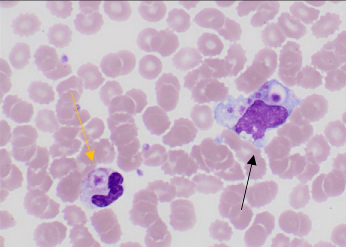
Figure 2:
Yellow arrow: Platelets within neutrophils
Black arrow: Platelets within monocytes

The patient was diagnosed with a community acquired pneumonia (CAP). Respiratory cultures did not yield any organism due to significant oropharyngeal contamination. He was started on a course of antibiotics with good response. Together with the clinical improvement of the patient, upon review of subsequent blood smears, there was resolution of leukocytosis and neutrophilic and monocytic engulfment of platelets.

This phagocytic phenomenon is rarely seen in the peripheral blood. This is likely a result of the release of pore-forming toxins from the pneumonia, leading to platelet activation to trap CAP pathogens1,2,3. Activated platelets are subsequently cleared by neutrophils and monocytes, resulting in the phagocytic activity demonstrated in the pictures above. With resolution of the CAP and hence inflammation, the phagocytic activity resolves as well.

References

  1. Amouroux, I., & Lesesve, J. F. (2020). Neutrophilic thrombophagocytosis. Morphologie, 104(344), 73–75.https://doi.org/10.1016/j.morpho.2019.08.004
  2. Feldman, C., & Anderson, R. (2020). Platelets and Their Role in the Pathogenesis ofCardiovascular Events in Patients With Community-Acquired Pneumonia. Frontiers in  Immunology, 11. https://doi.org/10.3389/fimmu.2020.577303  
  3. Maugeri, N., Patrizia Rovere-Querini, Evangelista, V., Covino, C., Capobianco, A., Bellone, M., Piccoli, A., Licia Totani, Cianflone, D., Attilio Maseri, & Manfredi, A. A. (2009). Neutrophils phagocytose activated platelets in vivo: a phosphatidylserine, Pselectin, and β2 integrin–dependent cell clearance program. American Society of  Hematology, 113(21), 5254–5265. https://doi.org/10.1182/blood-2008-09-180794  
TOP