Hassan MM1, Trina SS2, Ahmedullah AKM3, Islam MN4
¹Assistant Professor, Department of Medicine, Bangabandhu Sheikh Mujib Medical University, Shahbag, Dhaka-1000, Bangladesh.
²Registrar and rheumatologist, Department of Medicine, Sylhet MAG Osmany Medical College, Bangladesh.
³Associate professor, Department of Rheumatology, Bangabandhu Sheikh Mujib Medical University, Shahbagh, Dhaka-1000, Bangladesh.
⁴Professor of rheumatology, Department of Rheumatology, Bangabandhu Sheikh Mujib Medical University, Shahbagh, Dhaka-1000, Bangladesh.
*Corresponding author
*Hassan MM , Assistant Professor, Department of Medicine, Bangabandhu Sheikh Mujib Medical University, Shahbag, Dhaka-1000, Bangladesh.
Abstract
Background: Behcet’s disease (BD) is not common In Bangladesh. The diagnosis of BD is usually delayed also. Here, a Bangladeshi girl took six years to be diagnosed as a case of BD. She suffered from recurrent attack of fever with some unexplained rare symptoms and diagnosed as a case of BD.
Case summary - A 16-year-old girl suffered from recurrent episodes of fever, itch and joint pain and treated as viral arthritis. After three years of suffering, she was presented with periorbital edema, tender migrating rash, and myalgia. Then she went to neighboring country, where she was discharged with the diagnosis of adult-onset Still’s disease. At that time, she had fever, arthralgia and rash. Her RA, anti-CCP, ANA was negative but ferritin was raised and perivascular lymphocyte infiltration was seen in skin biopsy. She was put on prednisolone and methotrexate and She had partial improvement. She stopped medication after seven months except steroid, which she took during symptoms. After six years of illness, she was again presented with periorbital edema, migrating tender rash, red eye (scleritis) and swollen fingers (dactylitis). Then she got admitted in Bangabandhu Sheikh Mujib Medical University. She had hepato-splenomegaly, mild portal hypertension, gastropathy and portal fibrosis with few septa. Her skin biopsy showed leucocytoclastic vasculitis. With the diverse features, she was examined thoroughly and a few small ulcers was found in the lower lip. She also mentioned about recurrent aphthous ulcers with spontaneous healing. She also had occasional small genital ulcers that healed spontaneously. With all the features, Behcet’s disease was considered as a possibility and her pathergy test was positive and her HLA-B52 was positive but HLA-B51 was negative.
Conclusion – In rare diseases, it is difficult to diagnose if distinct features are absent. In that case milder or nonspecific features need adequate attention for early diagnosis. Here, mild aphthous and genital ulcers were important for early diagnosis of this multi-faced presentation of Behcet’s disease.
Key words: Behcet’s disease, apthous ulcer, HLA-B52
Introduction
Behcet’s disease can involve any organ of the body. It usually presents with oro-genital ulcer with or without ocular, cutaneous, articular, vascular, neurologic and other organ involvement 1. BD is thought to be an ancient silk route disease but has a worldwide distribution 2. The prevalence in Asian region was estimated 2.1-19.5/100,000 and also reported in southern and northern Europe 3-4. It is prevalent in Turkey followed by Iran, Israel and Japan but it is rare in Australia, America and Africa 5-6. Onset is usually in the 2nd to 4th decade but rare in children and elderly 7, 8. HLAB51 positivity is associated with the development of BD 9. There is no significant male/female distribution but in America BD is commonly reported in female 10. In Bangladesh, a few cases were reported 11.
Atypical features such as optic neuritis, dural sinus thrombosis, peripheral neuropathy, aseptic meningitis, brain stem or corticospinal tract symptoms, cerebellar symptoms, deep vein thrombosis, Budd-Chiari syndrome, pulmonary artery aneurysm, myocardial infarction, superior and inferior venacaval obstruction were reported previously 1. Sometimes, mild typical features are ignored or missed, and it is also difficult to diagnose a disease that presents with atypically features. This patient developed mild oro-genital symptoms after three years of sufferings. She did not mentioned it and it was retrieved later. She suffered from recurrent bouts of fever, rash, arthralgia and it was managed symptomatically. But, subsequent development of new features and evaluation by physician of home and abroad failed to solve her problem. After six years from the onset of her symptoms, she developed new features. And she admitted in a tertiary care hospital for further evaluation. The appearances of new features like urticaria, scleritis, dactylitis, hepatosplenomegaly is not considered as a typical feature of Behcet’s disease. She was examined properly and a few tiny apthous ulcer was seen in her lower lip. She also admits that she had this type of mild apthous ulcers in the past that was healed spontaneously. She also mentioned that she had small genital ulcers in the past that she did not realize a real problem. But these findings were the key features in diagnosing a rare disease like BD.
Case presentation
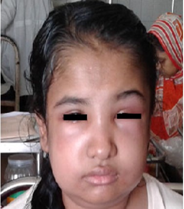
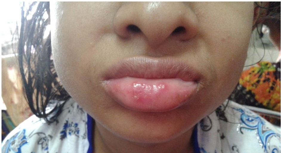
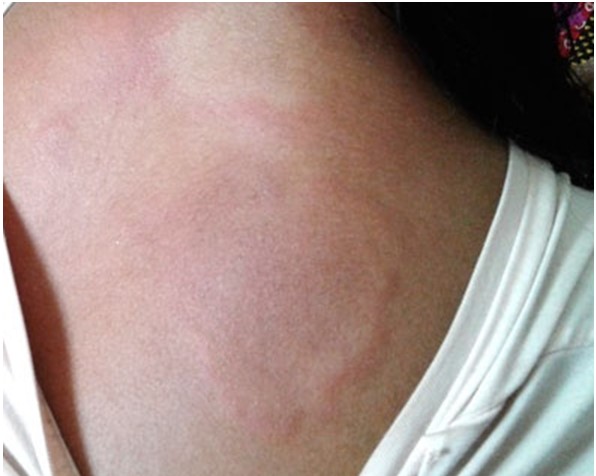
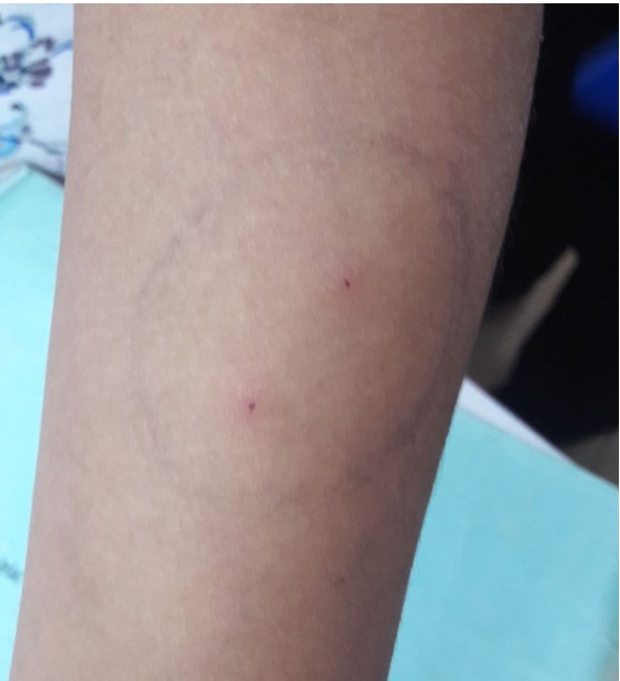
A 16-yr-old Bangladeshi girl suffered from a long course of illness. Six years back she had fever, arthritis and rash. At that time, she was treated as viral arthritis and symptoms were subsided after 3 to 4 days. But she experienced similar attack several times in the subsequent years, and attacks were triggered by minor physical stress and sometimes spontaneously. Interval between attacks varied between 1-8 weeks and occurred approximately 5 times per year. Her highest asymptomatic period was 3 months. She consulted several physicians and failed to reach any conclusion. She went to neighboring India for better management. At that time, she was also suffering from pain in multiple joints involving knee, ankle, wrist, elbow and shoulder. She also had hepatosplenomegaly. She had fever and she had tender palpable erythematous rash (Fig-3) also. With a periodicity of the fever, she was evaluated for periodic fever. Tumor necrosis factor (TNF) receptor-1 associated periodic fever syndrome (TRAPS) was considered as a possibility on the basis of recurrent fever, arthritis, periorbital edema, tenderness over migrating rash and protracted myalgia with perivascular lymphocyte infiltration in skin biopsy. But in TRAPS, each febrile period usually last more than 5 days. Here, each episode persists for 3-4 days. There was no history of affected family member. TRAPS is caused by TNFRSF1A that could not be tested due to unavailability. The she was considered as a case of Still’s disease for fever and joint pain. Her rheumatoid factor and anti-nuclear factor were negative but ferritin was mildly raised (683 ng/ml). she had hepato-splenomegaly. Considering all the evidence, she was labelled as a case of Still’s disease. And, she was put on deflazacort and methotrexate. She took deflazacort for 7 months (36 mg for 6 wks, 30 mg for 10 wks, 24 mg for 4 weeks, 18 mg for 4weeks, 15mg for one week) and methotrexate 15mg weekly. After seven months of treatment, she did not achieved remission. During this period her frequency of fever, skin rash and arthritis was reduced but did not stop happening. Later, she started to take steroid during symptoms and stopped following physician advice and follow-up. After several months, she got admitted in Bangabandhu Sheikh Mujib Medical University with a new flare of fever, arthralgia, coarse tremor, oro-genital ulceration and multiple urticarial rashes with periorbital edema (Fig-1). Fever was high grade, intermittent, highest recorded temperature was 103°F and persisted for 3-4 days. She noticed migratory skin rashes involving face, neck, chest, arms, fingers and toes. Rash was tender and swollen and it persisted for 2-3 days after fever was subsided. She had protracted myalgia and arthralgia with fever. Her oral (Fig-2) and vulval ulcerations were small, occurred in crops of 3-4 lesions and healed spontaneously for last 3 years. They start as pruritic papule which was ulcerated and healed without scarring. But the ulcer were small and she did not recognize it as a problem. She is the 2nd issue of her non-consanguineous parents. There was no such family history.
Figure 1: Periorbital edema
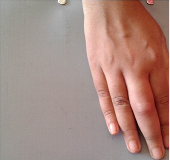
Figure 4: Dactylitis
She had erythematosus rash with puffiness over upper eyelid. Her temperature was 101°F, pulse was 110/min, blood pressure 100/60 mm of Hg, respiratory rate was 16 breaths/min. There were 2-3 minor apthous ulcers over lower lip having whitish base with erythematous border.
There were two large raised annular erythematous rashes with serpiginous margin resembling urticaria, one with central clearing (Fig-3). There were also multiple rounded erythematous plaques like lesions over upper limbs and back of torso. These lesions were warm and tender to touch.
Liver was palpable, 5 cm from costal margin with upper border of liver dullness was in the right 5th intercostal space along the right midclavicular line. Surface of the liver was smooth, nontender, firm in consistency, no hepatic rub or bruit. There was grade II tenderness in PIPs of ring fingers of both hands and MCPs of right hand but there was no swelling or restriction of movement. Her middle finger was tender and swollen like sausage resembling dactylitis (Fig-4). She had scleritis in her right eye.
She was investigated extensively both home and abroad. Her CBC, Hb%, PBF were normal as well as renal and hepatic functions. Her ESR was 30 mm in 1st hour and CRP was high (12.5 mg/L). Chest X-ray was normal, USG showed enlarged Liver (16.9cm) with homogenous echotexture, focal hyperechoeic area seen anterior to porta-suggestive of focal fatty infiltration, Spleen is mildly enlarged (13.3 cm). MRI of brain was normal. Hepatic fibroscan - 7.9 kpa (F2) showed portal fibrosis with few septa. Mild portal hypertensive gastropathy was reported in endoscopy. Her serum albumin was low (28.62 g/L), LDH was mildly raised (752 U/L) on one occasion but later it reduced to normal, serum ferritin was raised (683.4 ng/ml, 759.70 ng/ml), serum IgE was high (691 U/ml, >3000.0 IU/ml), serum TSH, FT3, FT4, S. ceruloplasmin, S. CPK and reticulocyte count was within normal limit. Malaria, kalazar and tuberculosis screening were negative. HBsAg, Anti-HBc (total), Anti-HCV were negative, VDRL and TPHA were nonreactive, skin smears for AFB and mycobacterial culture were negative as well as negative urine and blood culture. Her serum IgG, IgA, IgM levels were normal.
Figure 5: Pathergy test
ANA, Anti SSA, Anti SSB, C-ANCA, P-ANCA, Anti CCP were negative and serum C3, C4, were normal. Her Hb electrophoresis was Normal. Her skin biopsy revealed leucocytoclastic vasculitis evidenced by mildly dilated capillaries in the superficial dermis with unremarkable epidermis, moderate perivascular infiltrate of lymphocytes and polymorphs, Karyorrhectic debris and extravasated RBCs were seen. Endothelial cells were plumped. Doppler study showed caliber of portal vein was normal (8.2mm) with normal Doppler characteristics without any thrombus, flow direction is hepatorenal, peak systolic velocity is 21.5 cm/sec, mean velocity is 11.9 cm/sec, suggesting mild portal hypertension. There was mild tricuspid regurgitation seen in echocardiography. Caliber of hepatic vein was normal (3.9mm). Lumens are patent with hepato-fugal flow direction with typical Doppler spectrum. Diameter of inferior vena cava is 13.2 mm with patent lumen and there was no evidence of thrombus.
Previously Still’s disease was considered but diagnosis was excluded later because it didn’t fulfill the criteria and it is a diagnosis of exclusion. The duration of fever was short, typical salmon peak rash was absent, total WBC count is always less than 10,000, ferritin level was not markedly raised (<1000). Juvenile systemic lupus erythematosus was excluded as because she does not have hard palate ulcer, butterfly rash, photosensitivity, renal involvement and ANA was negative. She had erythematous upper eye lid swelling but did not have proximal weakness and her serum CPK, ALT and AST were normal. She had recurrent febrile episode but duration was not characteristic of familial autoinflammatory syndrome. TRAPS usually persist at least for 5 days, and presence of aphthous ulcer is a negative predictor for TRAPS (odds ratio 0.2) for periodic fever 12. Finally, small apthous ulcer was the new findings for which Pathergy test was done which turned positive (Fig: 5). And HLA-B51 was done and that came negative. Later HLA-B52 was advised and it was positive.
Her skin biopsy showed leucocytoclastic vasculitis. She scored four points labelled according to Revised International Criteria for Behcet’s Disease. As per Behcet’s Current Clinical Activity Form her disease activity index is seven. This case has many atypical manifestations which include urticaria like rash, hepatosplenomegaly, scleritis, portal hypertension, dactylitis and uncommon genotypic profile is the center point of discussion.
Discussion
As there is no confirmatory test, BD diagnosis is made mostly on the clinical criterion. Usually, BD is a chronic disease and its course is unpredictable. High index of suspicion and previous experience is necessary to reach the diagnosis. Diagnostic is delay is common 2 as nonspecific constitutional features predominate early and typical features appear later like this case. In this patient, initially it was thought that she had viral arthritis but recurrence of similar symptoms considered Still’s disease. But criteria was not fully matched for still’s disease. As total count was repeatedly normal and rash was not typical, serum ferritin was minimally raised and urticaria is a very unusual cutaneous manifestation in adult Still’s disease 13, 14 as well as aphthous ulcer has not yet been reported in still’s disease. For that other possibilities were reconsidered. Prevalence of aphthous stomatitis in Behcet’s is near 100% 15. TRAPS was a strong consideration with the presence of recurrent fever, migratory tender rash, protracted myalgia, periorbital edema. Although arthralgia is common but arthritis (1.2%), urticarial plaque (23%), aphthous stomatitis (13%), splenomegaly (11%), hepatomegaly (9%) has been reported in TRAPS 16. Here each febrile episode is 3-4 days but in TRAPS fever usually persists for 1-2 weeks 16. Which is an important point against TRAPS. Later TRAPS was challenged with the absence of family history, short duration of fever (< five days), presence of rash in different parts of the body and presence of recurrent vulval ulcer are not consistant with TRAPS. Exact diagnosis can only be made through genetic analysis to see mutation in TNFRSF1A 17. TRAPS is an uncommon disease and prevalence is one in a million 18. Many typical features were absent to consider both Still’s disease and TRAPS. Minor aphthous and genital ulcer were absent in first three years of disease coarse and then it was ignored both by patient and previous physicians. And her diagnosis was delayed for three more years.
When patient presented here, oral and vulval ulceration came to the cornerstone of evaluation and Behcet’s came to the spotlight. But hepatosplenomegaly, urticaria, scleritis and dactylitis were not common in BD. This patient also presented with recurrent mild short bout of fever not common in Behcet’s though BD is a cause of pyrexia of unknown origin 19. Fever is more common in BD with vascular involvement than with muco-cutaneous lesions 20. In a study, 22% BD patients had fever with vascular, neurologic or joint involvement 21. Her skin biopsy showed leucocytoclastic vasculitis which can be caused by Behcets (17%) 22. Patients with positive HLAB51 may exhibit arthritis and urticaria like syndrome 23, but she is HLAB52 positive. Uveitis is characteristics of BD, but scleritis even nodular pattern has association with BD 24. She had coarse action tremor which can also be explained with Behcet’s as it occurs in 6% of cases 25. The patient has dactylitis, that is not feature of BD. In a case report, BD with undifferentiated spondyloarthropathy have dactylitis that later developed psoriatic plaque after a follow up of 3yrs 26. She had another atypical feature hepato-splenomegaly, which is rare in BD. One explanation could be nodular regenerative hyperplasia of liver progress to non-cirrhotic portal hypertension that was reported previously 27. HLAB51 is a known genetic risk factor for Behcet’s, but here she is HLA-B52 positive. In Iranian, Azari 29 and Israeli 28 patients with BD has shown HLA-B52 positivity. It is also found to be positive in Behcet’s disease having multi-arterial lesions 30.
BD is a chronic and typically self-limiting inflammatory condition. Severity of flares is unpredictably ranges from mild to severe. Frequency and duration of illness does not follow any fixed pattern 31. This patient had a wax and waning course of clinical illness with many atypical findings before her diagnosis was made.
References
- Kaufman S W, McNamara EK, Jorizzo JL. Behcet’s disease: In Firestein GS, Budd RC, Gabriel SE, McInnes IB and O’Dell JR editors. Kelley and Firestein’s Textbook of Rheumatology. 10th ed. Philadelphia PA: Elsevier; 2017. P. 1589-1596.
- Nieves Marie Leonardo and Julian McNeil, “Behcet’s Disease: Is There Geographical Variation? A Review Far from the Silk Road,” International Journal of Rheumatology, vol. 2015, Article ID 945262, 7 pages, 2015. https://doi.org/10.1155/2015/945262.
- Mahr A, Maldini C. Epidemiology of Behcet’s disease. Rev Med Interne. 2014; vol 35(2):81-89.
- Mohammad A,Mandl T,Sturfelt G,Segelmark M. Incidence, prevalence and clinical characteristics of Behcet’sdisease in Southern Sweden. Rheumatology.2013; vol.52(2):304-310.
- Wakefiled D and McCluskey P.Behcet’s syndrome: Ocular features in Australian population. Australia and New Zealand Journal of Opthalmology.1990; vol.18(2): 129-135.
- Keino H and Okada A. Behcet’s disease: Global epidemiology of an old Silk Road disease. British Journal of Opthalmology.2007;vol.91: 1572-1574.
- Saadoun D and Wechsler B. Behcet’s disease. Orphanet Journal of Rare diseases.2012;vol.7(1).
- Davatchi F ,Shahram F , Davatchi CC, et al. Behcet’s disease: from east to west. Clinical Rheumatology.2010; vol.29(8) :823-833.
- deMenthon M, Lavallery MP, Maldini C, et al. HLAB51/B5 and the risk of Behcet’ s disease; a systematic review and meta analysis of case-control genetic association studies. Arthritis Rheum.2009;vol.61:1287.
- Sibley C, Yaziciy Y, Tascilar K, et al. Behcet’s syndrome manifestations and activity in the United States versus Turkey-A cross sectional cohort comparison. J Rheumatology.2014;vol.41(7): 1379-1384.
- Samad T, Ahmed JU, Rahim MA, et al; Behcet’s disease presenting as fever of unknown origin:A case report. Birdem Medical journal.2013; vol.3(1).
- Gattorno M, Sormani MP, DOsualdo A, et al. A diagnostic score for molecular analysis of hereditary autoinflammmatory syndrome with periodic fever in children. Arthrits Rheum. 2008;vol.58:823.
- Bas Y, Sechin YH,Kalkan G, et al. Investigation of Behcet’s disease and recurrent apthous stomatitis frequency: The highest prevalence in Turkey. Balkan Med J. 2016;33:390-5.
- Salaffi F, Filosa G, Bugatti L, Maestrini MD. Urticaria as a presenting manifestation of adult- onset Still’s disease. Clin Rheumatol.2000;19(5):389-9.
- Criado RF, Criado RR, Vasconcellos C, et al. Urticaria as cutaneous sign of adult-onset Still’s disease. J Cutan Med Surg. 2006;10(2):99-103.
- Lachmann H J, Papa R, Gerhold K, et al. The phenotype of TNF receptor associated autoinflammatory syndrome (TRAPS) at presentation: a series of 158 cases from the EUROfever/EUROTRAPS international registry. Ann Rheum Dis.2014; 73: 2160-2167.
- Aksentijevich I, Galon J, Soares M, et al. The Tumor-Necrosis-factor Receptor- Associated Periodic Syndrome: New mutations in TNFRSF1A, ancestral origins, genotype-phenotype studies, and evidence for further genetic heterogenecity of periodic fevers. Am. J.Hum.Genet.2001; 69:301-314.
- Lainka E, Neudorf U, Lohse P, et al. Incidence of TNFRSF1A mutations in German children; epidemiological, clinical and genetic characteristics. Rheumatology.2009;48:987-991.
- Saltoglu N, Tasova Y, Midikli D, Aksu S.ZH, Sanli A, Dundar HI. Fever of unknown origin in Turkey; evaluation of 87 cases during a nine-year-period of study. Journal of Infection. 2004;vol.48(1):81-85.
- Hatemi G, Seyahi E ,Fresho I, Talarico R, Hauryudan V; Behcet’s syndrome : A critical digest of the 2013- 2014 literature. Clin Exp. Rheumatology.2014. vol. 32 (suppl 84).
- Seyahi E, Karaaslam H, Ugurlus S, Yazici H. Fever in Behcet’s syndrome. Ciln Exp Rheumatol.2013;31(3 suppl 77): 64-7.
- Chen KR, Kawahare Y, Miyakawa s. Mishikua T. Cutaneous vasculitis in Behcet’s disease: A clinical and histopathological study of 20 patients. Journal of the American Academy of dermatology.1997; vol.36(56): 689-696.
- Pasero G, Olivieri I, Gemignani S, Vitali C. Urticaria/arthritis syndrome: report of 4 B51 positive patients. Annals of Rheumatic diseases.1989; vol.48: 508-511.
- Chang KH and Cho HE.A case of nodular scleritis in association with Behcet’s disease. The Korean Journal of Internal Medicine. 2001;vol.1(1).
- Behcet’s syndrome : Pathogenesis, clinical manifestations and treatment. Seyahi E, Hatemi G, Fresho I, Melikoglu M, Yazici H. Oxford Textbook of Vasculitis, 475, 3rd edition.
- Padula A, Ciancio G, Cantini F,et al. Coexisting Behcet’s syndrome and Spondyloarthritis. Clin Rheumatol. 1999; 18(6):499-500.
- Bloxham CA, Henderson DC, Hampson J, Burt AD. Nodular regenerative hyperplasia of Liver in Behcet’s disease. Histopathology. 1992;vol.20(5): 452-454.
- Arber N, Klein T, Meimer Z, Pras E, Weinberger A. Close association of HLAB51 and B52 in Israeli patients with Behect’s syndrome. Annals of Rheumatic Disease.1991; 50: 351-355.
- Shahneh ZF, Hamzavi F, Bayazi B, Banbelhagh A, Baradaran B. New insights into HLA class I association to Behcet’s syndrome in Iranian Azari patients. Autoimmun Highlights.2013; 4: 101-102.
- Sugisaki K, Saito R, Takagi T,et al. HLAB-52-positive vasculo-Behcet disease: Usefulness of magnetic resonance angiography, ultrasound study, and computed tomographic angiography for the early evaluation of multiarterial lesions. Mod Rheumatol.2005; 15(1):56-61.
- Zeiden JM, Saadaun D, Garrido M, Klatzmann D, Six A, Cocouh P. Behcet’s disease pathophysiology: A contemporary review. Autoimmun Highlights.2016; 7:4.