
Numerous Ectopic Sebaceous Glands in the Esophagus; A Case Report and Assumption regarding Pathogenesis
Masao Suzuki1,4, Shoko Kawamura2, Hideki Mori3*
1Department of Gastroenterology/Endoscopy Center, Ogaki Tokusyukai Hospital, Japan
2Medical Checkup Center, Ogaki Tokusyukai Hospital, Japan
3Department of Diagnostic Pathology, Ogaki Tokusyukai Hospital, Japan
4Present address: Division of Gastroenterology, Juko Kinen Hospital, Japan
*Corresponding author
Hideki Mori, Department of Diagnostic Pathology, Ogaki Tokusyukai Hospital, Emeritus Professor Gifu
University
DOI: 10.55920/JCRMHS.2023.04.001162
Abstract
It is known that ectopic sebaceous glands (ESG) occur infrequently in the esophagus. ESG with numerous lesions of the esophagus are extremely rare. An endoscopic examination of a 79-year-old man revealed the presence of ESG more than 100 tiny lesions scattered throughout the esophagus. Immunohistochemically, keratin 14 (cytokeratin K14), ER (estrogen receptor) and Ki67 were predominantly expressed in the basal cells of ESG. Meanwhile, GATA3 (zinc finger transcription factor) was recognized mainly in the sebocytes of them. Clinicopathology of ESG and an assumption regarding pathogenesis of ESG, particularly in view of acquired metaplasia versus congenital misplacement is stated.
Introduction
Sebaceous gland is closely related to the hair follicle, forming the pilosebaceous apparatus, most commonly in face and scalp, but occasionally in aberrant areas. Ectopic sebaceous glands (ESG) have been found in organs of ectodermal origin, such as lips, oral cavity and parotid glands. ESG in esophagus are rare, since the organ is endodermal-derived tissue. The first report of gross findings of ESG was based on the autopsies in 1962, which revealed ESG in approximately 2% of the totals (1). Since then, less than 50 cases have been identified and presented in the medical literature (2). For the advance of endoscopic screening, cases of ESG have been incidentally found during routine upper endoscopy for other reasons (2, 3). Presence of lobules of cells with sebaceous differentiation in the lamina propria and proliferating basal cells within the epithelium are characteristic findings of ESG.
In several studies, esophageal ESG were followed up for years, but no changes in the number and size of the lesions were eventually observed (4-6). Furthermore, no reports on malignant transformation have been elucidated. Importantly, a few cases with numerous ESG with high number more than 100 lesions have been manifested (7-11). Peculiarly, these cases were found preferably in Asian countries. Recently, we encountered a case with numerous ESG (more than 100) throughout the esophagus. An assumption regarding the pathogenesis as well as clinicopathology of the case is described.
Case report
A 79-year-old man complaining of heartburn was referred to our hospital for endoscopic examination. The patient had smoked about 15 cigarettes daily and had been a moderate rice wine drinker. His family history was unremarkable. The patient had weak anemia (erythrocytes 392x104/μl, hematocrit 35.9%). His serum cholesterol level was within normal range (188 mg/dl). An endoscopic examination revealed the presence of more than 100 tiny, rounded, elevated, yellowish lesions <0.5mm in diameter scattered throughout the upper, middle and lower esophagus (Figure 1). In some of the lesions, discharge of whitish fat like substances from ESG was observed.
Histologically, ESG of the esophagus was identified as mature sebaceous glands in lamina propria. Moderate lymphocytic infiltration was frequently recognized surrounding ESG (Figure 2). Immunohistochemically, k14 (cytokeratin 14) was strongly expressed in the ESG together with neighboring basal cells (Figure 3).
Furthermore, Ki 67 being a marker of cell proliferation was recognized in the basal cells of ESG as seen in the basal cells of squamous cell epithelium near by the ESG. Estrogen receptor (ER) and GATA3 (zinc finger transcription factor) were also expressed in the basal cells and sebocytes of ESG respectively (Figures 4, 5), although positive response of progesterone receptor (PGR) was not apparent.
In the esophagus of the patient, transverse mucosal break <5mm was detected. The esophagus of this patient was the state of Grade A reflux esophagitis (Los Angeles Classification). Barrett’s esophagus was not confirmed. For the stomach, was recognized scattered fundic gland polyps (1〜2mm). On the same day for the endoscopic examination, a polyp was found in the cecum, and was removed by the endoscopic mucosal resection. The polyp was later diagnosed as tubular adenoma accompanying focal adenocarcinoma. He is healthy and is under observation by medical checkup center of our hospital.

Figure 1: Macroscopic appearance of ESG in the esophagus. Multiple slightly elevated, yellowish lesions are seen.

Figures 2 : Histology of ESG. Mature sebaceous glands associated with moderate lymphocytic infiltration lie in the lamina propria. H&E stain
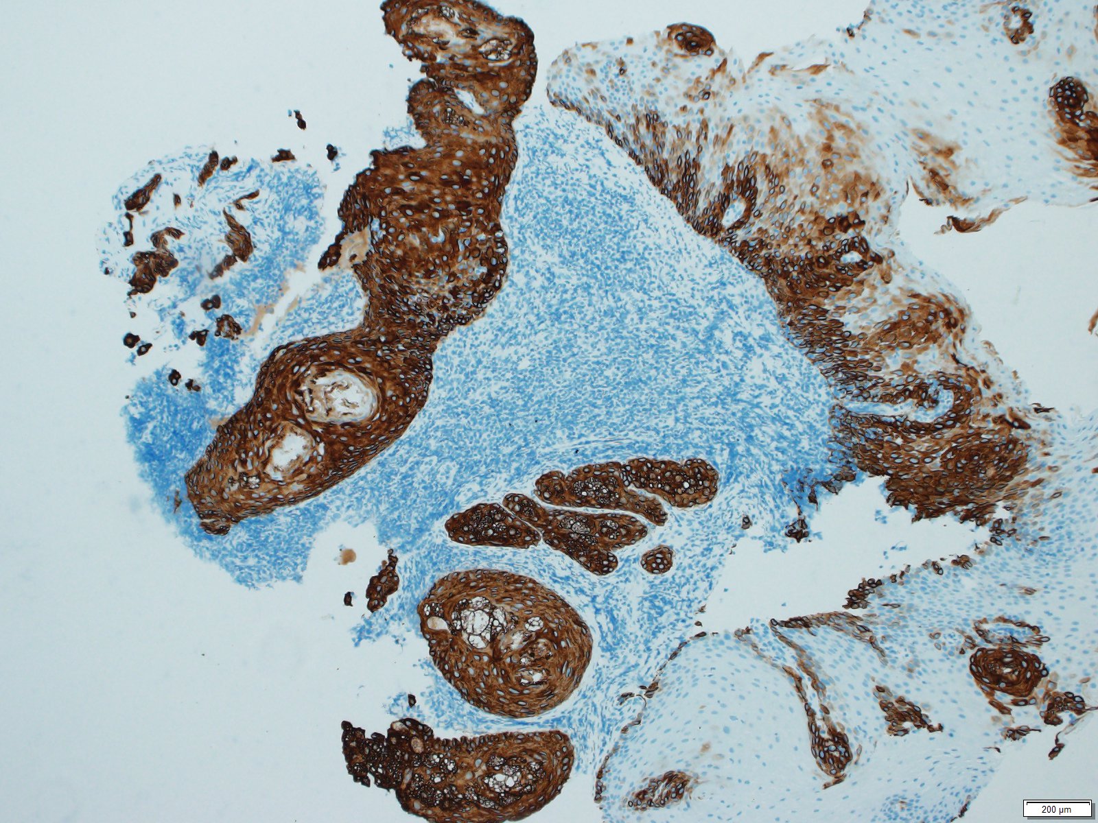
Figures 3:Histology of ESG. K14 is strongly expressed in the ESG and basal cells of neighboring squamous epithelium.

Figures 4 :Histology of ESG. ER is expressed in the basal cells of ESG.

Figures 5 :Histology of ESG. GATA is expressed in the sebocytes of ESG.
Discussion
The origin of ESG in the esophagus and the mechanism of their development are unclear. The most interesting aspect is that the esophagus originates from the endoderm, whereas the sebaceous gland is the structure of the ectodermal origin (3). It is postulated that the entity is an acquired metaplastic process or a congenital misplacement. Nakanishi et al (9) described that metaplasia of the basal cells toward sebaceous differentiation occurred by acquired stimulus like chronic inflammation on the bulbous nest of the basal cells. Thus, the metaplastic theory seems to be plausible. They examined to know the relationship between keratin expression and sebaceous differentiation using 6 different anti-keratin antibodies. As a result, only keratin 14 was demonstrated in bulbous nest of proliferating basal cells. In our study, expression of keratin 14 was also confirmed in the ESG and neighboring basal cells of the squamous epithelium. Thus, basal cells with expression keratin 14 may be importantly related to sebocyte differentiation. In our study, basal cells of ESG exerted higher labeling of Ki 67 suggesting potential activity of cellular growth of ESG. The evidence suggests that ESG survive longer under the rigorous environment of the esophagus.
Reflux esophagitis as shown in the present case is a representative chronic inflammation of esophagus, esophageal epithelium is exposed to gastric juice which consists of sodium chloride and pepsinogen. In some case of esophageal reflux, duodenal juice containing cholic acid and bilirubin is also concerned. It is also known that reflux of gastric and duodenal juice causes otitis media as well as laryngopharyngitis (12). Thus, it is reasonable that damaged squamous cell epithelium with consistent exposure of the toxic agents contained in the duodenal as well as gastric juice cause metaplasia of the basal cells toward sebaceous differentiation. It might be also possible that cytokines derived from proliferated lymphocytes promote cell growth and is related to the development of ESG, although no evidences are obtained.
On the other hand, Hashimoto et al (11) emphasize that ESG are of ectodermal origin, despite the digestive tract being of endodermal origin, thereby congenital misplacements under hormonal regulation should be important. They had an assay on large scale of immunohistochemistry for the expression of hormone receptors of the constituted cells in ESG. In their study, hormone receptors like androgen receptor, ER and PGR, and GATA being a transcription factor to regulate the differentiation of mammary glands (13), displayed positive responses in the sebocytes and basal/parabasal cells in different cases of ESG.
Present authors also surmise that histogenesis of ESG with numerous lesions may be different from that with a few lesions. Squamous cell epithelium of the patient having ESG with numerous lesions may already acquire a predisposition for the sebaceous differentiation leading to ESG. In this study, expression of GATA3 and ER was confirmed immunohistochemically in the sebocytes and basal cells of ESG respectively. Such evidences are in consistent with those shown by Hashimoto et al (11). Accordingly, development of ESG, particularly the type with numerous lesions may be attributable to both of congenital misplacement and acquired metaplasia.
Meanwhile, some of recent study reports that Hedgehog signaling pathway plays a key role for the development of sebocytes and might be a potential target for studying other similar disorders related to abnormal sebaceous gland function (14). At present, precise etiology of ESG of the esophageal epithelium remains to be determined. Further studies of esophageal ESG are required to manifest the causes and clinicopathological process of such unusual epithelial alteration.
References
- De la Pava S, Pickren JW, Ectopic sebaceous glands in the esophagus. Arch Pathol, 73, 397-399, 1962.
- Baffa C, Naaman A, Coppola D, Sebaceous gland ectopic of the esophagus: A challenging clinical diagnosis, In vivo, 36, 2923-2926, 2022.
- Jeon MS, Kim GH, Jeng DY et al, Magnifying endoscopy for esophageal ectopic sebaceous glands, Clin Endosc, 51, 495-497, 2018.
- Marcial MA, Villafana, Esophageal ectopic sebaceous glands: endoscopic and histologic findings, Gastrointest Endosc, 40, 630-632, 1994.
- Nishisaki H, Yasutake K, Nakashima T et al, Five cases with ectopic esophageal sevacecous glands, Dig Endosc, 9, 207-212, 1997.
- Fukuchi M, Tsukagoshi R, Sakurai S, et al, Ectopic sebaceous glands in the esophagus: endoscopic findings over three years, Case rep gastroenterol, 6, 217-222, 2012.
- Bertoni G, Sassatelli R, Nigrisoli E et al, Ectopic sebaceous glands in the esophagus: report of three cases and review of the literature, Am J Gastroent 89, 1884-1887, 1994.
- Hoshika K, Inoue S, Mizuno M et al, Endoscopic detection of ectopic multiple minute sebaceous glands in the esophagus. Report of a case and review of the literature, Dig Dis Sci, 40, 287-290, 1995.
- Nakanishi Y, Ochiai A, Shimoda T et al, Heterotopic sebaceous glands in the esophagus: histopathological and immunohistochemical study of a resected esophagus, Pathol Int., 49, 364-368, 1999.
- Wang WP, Wang WS, Tsai YC, Multiple tiny ectopic sebaceous glands discovered throughout entire esophageal tract, Dig Dis Sci 54, 2754-2757, 2009.
- Hashimoto H, Horiuchi H, Miura S et al, Clinicopatholologic characteristics of esophageal ectopic sebaceous glands: Chronological changes and immunohistochemical analysis, Int J Surg Pathol, 29, 378-384, 2021.
- Sone M, Yamamoto Y, Hayashi H et al, Otitis media in adults as a symptom of gastroesophageal reflux, Otolaryngol Head Neck Surg 136, 19-22, 2007.
- Bai F, Zhang LH, Liu X et al, GATA3 functions downstream of BRCA 1 to suppress EMT in breast cancer, Theranostics, 11, 8218-8233, 2012.
- Grachtchouk AM, Sheng H, Grachtchouk V et al, Hedghog signaling regulates sebaceous gland development, Am J Pathol, 163, 2173-2178, 2003.