What is the Sentence for Patients with High-Grade Gliomas: Tumor Structure or Mutation of H3F3A, IDH1, BRAF Genes?

O.S. Regentova1, O.I. Antonenko1, R.A., N.I. Zelinskaya1, R.A. Parkhomenko 1,2, Rogova T.S.1 A.I. Shevtsov1, M.A. Bliznichenko1, V.A. Solodkiy1

¹Russian Scientific Center of Roentgenoradiology (RSCRR), 86 Profsoyuznaya St., Moscow, 117997, Russia
²RUDN University, 6 Miklukho-Maklaya St., Moscow, 117198, Russia

*Corresponding author

Olga Regentova, Russian Scientific Center of Roentgenoradiology (RSCRR), 86 Profsoyuznaya St., Moscow, 117997, Russia
Email: olgagraudensh@mail.ru

Abstract

High grade gliomas (HGG) represent a serious problem for pediatric oncologists. Despite the improvement of the treatment of HGG over the past decades, its results are still far from satisfactory. The purpose of our study was to evaluate the prognostic significance of certain radiological, morphological, and molecular genetic characteristics of the verified brain gliomas on the basis of our own material. We analyzed the influence of some factors (tumor structure, volume of surgery, mutation status) on the overall survival in the cohort of 94 patients with HGG who received radiation or chemoradiotherapy at the Federal State Budgetary Scientific Center of Roentgenoradiology of the Ministry of Healthcare of the Russian Federation from 2016 to 2018. The best overall survival rates in the study group were observed in patients after radical surgery, in the absence of the K27M H3F3A gene mutation, in the predominance of a dense tumor component without necrosis zones according to MRI.

Keywords: gliomas in children, HGG, pediatric oncology, molecular genetic research, H3F3A K27M

Introduction

According to the literature data of the last 20 years, brain tumors in children aged 1 to 19 years consistently occupy the second place among the causes of death from malignant tumors after hemoblastoses. While significant progress has been made in the treatment of a number of other cancers during this period, there are few changes in the results for brain tumors, especially gliomas [1]. One of the reasons for this situation is the molecular genetic heterogeneity of the cell population forming glial tumors. That heterogeneity leads to phenotypic variability at the cellular level, providing multiple mechanisms of resistance to existing treatments for the disease. The intratumoral heterogeneity of glioma and the plasticity of malignant cells represent huge obstacles to the development of targeted treatment methods [2]. In recent years, many studies have been devoted to the search for ways to personalize glioma treatment methods in adults, which, unfortunately, have not yet resulted in breakthrough results. In pediatric neurooncology, the results are even more modest, since the number of clinical observations in this age group is significantly less than in the adult population. Moreover, as has already been established, molecular structure of high-grade gliomas in children differs from that of adults and, in fact, even in the presence of common histological and immunohistochemical features, they have specific genetic and epigenetic changes [3]. The need to identify molecular genetic subspecies of brain tumors is emphasized in the updated fourth edition of the World Health Organization (WHO) classification of tumors of the central nervous system (CNS) (2016). The integration of molecular and genomic features into histology has become fundamental in making diagnoses in the fifth edition of the WHO classification of CNS tumors (2021) [4]. As for pediatric CNS tumors, this has led to some changes in the classification of 2021: now there are families of tumors of the "pediatric type" and "adult type" for low- and high-grade gliomas. The molecular parameters were integrated into the new tumor classification, which is the result of a combined histological and molecular grouping within the tumor type. Thus, accurate molecular diagnostic methods are necessary for the personalized treatment of the diverse tumor variants [4]. Many researchers are currently exploring the possibility of using the results of the analysis of the histological and molecular genetic profile of the tumor to personalize the treatment of such patients [6]. Mutations in the H3F3A, BRAF, IDH1/2, TP53, and EGR genes, as well as the methylation status of the MGMT gene, are considered promising prognostic markers of glial tumors. An important finding in pediatric neuro-oncology in recent years has been the identification of histone H3F3A K27M (H3K27M) mutations in high-grade gliomas and the subsequent detection of this mutation in tumors of the midline structures of the brain, characterized by an extremely aggressive clinical course and an unfavorable prognosis [7,8]. Studies in this area has led to the identification of a separate variant of diffuse midline K27M mutant glioma, a highly malignant tumor (grade 4) with predominantly astrocytic differentiation. Diffuse gliomas with the K27M mutation are most commonly localized in the midline structures of the brain. For the first time, this variant of glioma was allocated to a separate category in the WHO classification of CNS tumors in 2016 [9]. The frequency of the K27M mutation, according to various researchers, is up to 50% in diffuse gliomas of the brainstem and in gliomas of the thalamus in children [10]. The main features of tumors with the K27M mutation in the H3F3A gene in children are the high tendency to progression and resistance to therapy, as well as the ability of tumor cells to migrate and invade [11].

A significant achievement in neurooncology was the study of the BRAF protooncogene from the family of RAF genes (BRAF, BRAF and CRAFT). The BRAF V600E mutation is most characteristic of low-grade gliomas (LGG) in children. The identification of mutations in the BRAF gene led to the development of antibodies specific to the V600E mutation, allowing for personalized targeted therapy [12].

Mutations in the IDH1 and IDH2 genes turned out to be favorable molecular genetic prognostic signs in adult gliomas. They are more common in benign forms of gliomas [15]. Mutations of the IDH1/2 genes in gliomas in children are detected in rare cases. At the same time, Pollack I. et al. noted that in children over 14 years of age, the IDH1 R132H mutation occurs with a frequency of up to 16% and is associated with a favorable prognosis [13]. The diagnostic and prognostic value of other molecular genetic anomalies was demonstrated, which were also included in the modern classification.  Those molecular genetic markers are widely used in the routine practice, being an integral part of the final diagnosis, as well as determining patient treatment tactics [14]. Today, it is already an axiom that a deep understanding of the molecular landscape of brain tumors in children and its role both in malignant transformation and in the development of therapeutic resistance, is important for personalization and improvement of both the established and proposed treatment methods [15].

In addition to the molecular and genetic characteristics of tumors, correlations of their prognosis with some clinical and radiological signs have become clearer. As in the adult population, the degree of radical surgical intervention affects the disease-free and overall survival of children with gliomas [5]. The evolution of brain tumor surgery, including that for deep-seated structures, has undergone major changes over the past 40 years. This is due to the emergence and improvement of microsurgical techniques and instruments, neuroimaging methods and programs, including magnetic resonance imaging (MRI), magnetic resonance tractography (MR-tractography), which make it possible to accurately determine the topographic and morphological features of tumors and their relationship to the functionally significant areas. At the same time, the rationale of resection of infiltrative tumors of deep localization remains controversial [16]. In some cases, the tumor fragments obtained during stereotactic biopsy of brain stem tumors, turn out to be too small, which creates certain difficulties in making a correct histological diagnosis [17].

The possible association between imaging characteristics of a pediatric brain tumor and the prognosis has not yet been studied sufficiently. On MRI images, glioma is usually defined as a mass of a dense or uneven structure, hypointensive on T1-weighted images and hyperintensive in T2 and FLAIR sequences [18]. There are a number of additional signs indirectly indicating the suspected degree of malignancy, such as the presence of cysts, necrotic zones, calcinates, invasion into neighboring structures, the presence of metastases and hydrocephalus [18,19]. A comprehensive assessment of the informative value of all these radiological symptoms in children, as well as molecular and genetic signs, seems necessary on the path of personalization of treatment methods. Considering the above, it seems appropriate to study the association between the most important molecular and genetic parameters of the tumor and some radiological and morphological characteristics in pediatric HGG.

Material and methods

We studied the long-term treatment outcomes of 94 children with HGG who received radiation or chemoradiotherapy at the Federal State Budgetary Institution of the Russian Federation for the Ministry of Health in the period from 2016 to 2018. The study included children and adolescents with supratentorial and infratentorial tumors, with the exception of diffuse brain stem tumors. In all patients, the diagnosis was verified by histological examination of the material obtained as a result of surgery or biopsy of the tumor. The median follow-up was 18 months. Mutations in the H3F3A, BRAF, and IDH1 genes were studied using Sanger sequencing in the material obtained during surgery. According to the initial MRI performed in various modes before surgery, the radiological structure of the tumor and the presence of destruction zones were evaluated. The volume of surgical intervention was assessed on the basis of the surgery protocols, the degree of malignancy was defined during the histological examination of the material obtained as a result of surgery or biopsy. The main criterion for the effectiveness of treatment was the life expectancy of children, with the starting point being the first day of treatment and the end point being the date of the last observation or death (Table 1).

All patients in the analyzed group underwent radiation therapy to the initial lesion area up to the total dose recommended by the clinical guidelines of the Ministry of Health of the Russian Federation for the treatment of CNS tumors in children [20]. Radiation therapy was performed using a 3D conformal technique after 3D planning (IMRT/ VMAT), in the standard fractionation regimen with single dose of 1.8-2.0 Gy up to total dose of 54 Gy in children younger than 5 years old and up to 60 Gy in older patients. Radiation therapy was combined with concurrent chemotherapy with temozolomide 75 mg/m2 daily. Depending on the clinical situation, in order to prevent and correct radiation reactions, RT was performed simultaneously with dexamethasone 0.15 mg/kg daily, and acetazolamide in an age-related dosage regimen three days a week, while PPIs, potassium-sparing drugs, and antifungal therapy were used in parallel.

Statistical data processing was carried out with the Statistica 13 program. Actuarial overall survival was calculated using the Kaplan-Meyer method.

Results

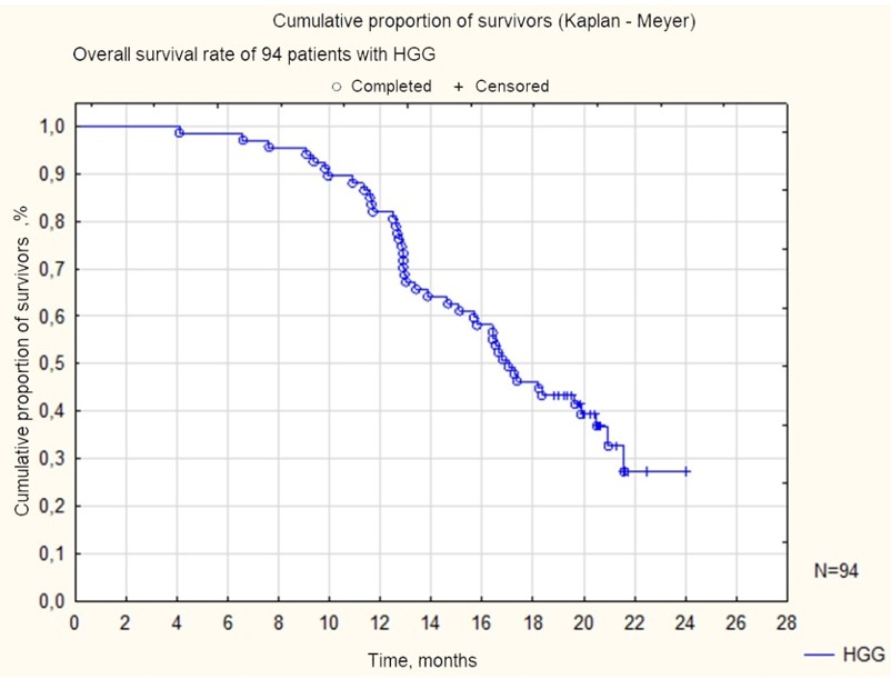
The diagnosis and monitoring of the tumor process in neuro-oncology is mainly based on the analysis of morphological and radiological methods. At the same time, there are practical difficulties, such as the subjectivity of the assessment of radiological and microscopic images, as well as the lack of the possibility of personalized therapy with a more detailed analysis of the disease during and after treatment. The results of long-term study have not brought significant changes for this group of patients today. As expected, all 94 patients with high grade gliomas treated at our Center had a predicted the median of survival rate up to 24 months, and only half of the children lived to 18 months (Fig. 1).

Figure 1: The overall survival rate of 94 children with high-grade gliomas.

According to the results of the study, we did not find a statistically significant effect of gender and age on OS for the entire research group.

Early diagnosis of gliomas determines the success of treatment and, accordingly, affects the prognosis. Therefore, the use of various medical imaging techniques is necessary to make the right decision when choosing therapeutic tactics, those techniques include MRI. MRI digital images of a tumor reflect its anatomical and functional changes, that is why MRI is the method of choice. A typical picture of a high-grade glioma looks like the lesion of a dense or uneven structure; the growth pattern may be solid, cystic or diffuse. There are many additional features that indirectly reflect the grade of glioma, such as the presence of cysts, necrotic zones, and calcinates - also reflect the level of malignancy. It can be noted that the solid structure of the tumor on the initial MRI in our cohort was a favorable prognostic sign. The predicted OS for up to 24 months for the patients with such tumors was more than 40%, while in the presence of signs of disintegration (cystic component) of the HGG all patients died within 18 months (p=0.000) (Fig. 2).
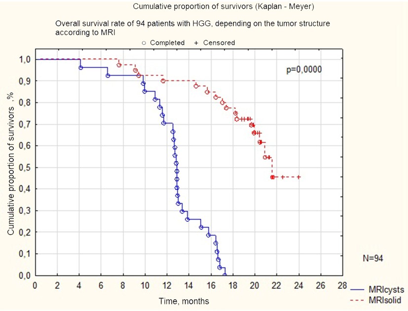
Figure 2: The overall survival rate of patients with high-grade gliomas depending on the structure of the primary tumor according to primary MRI of the brain.

The mutational load of a CNS tumor is often associated with a worse OS. However, not all mutations of genes specific to brain gliomas are associated with the same prospects. In our study, the main mutations of potential prognostic significance were studied, such as the K27M mutation in the H3F3A gene, V600 in the BRAF gene and R132H in the IDH1 gene. The fact of the presence or absence of changes in the above-described genes turned out to be fundamental for the prognosis of the entire analyzed cohort of children with HGG. The absence of those gene changes correlated with an increase of OS. More than 30% of the analyzed patients who had no gene changes survived the two-year period (Fig. 3).
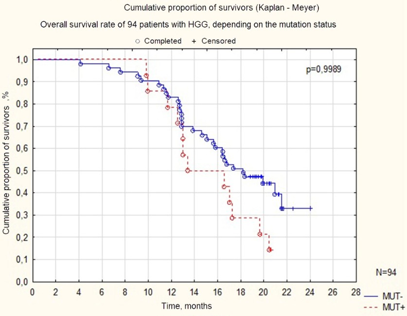
Figure 3: Overall survival of all patients with HGG, depending on the mutation status.

The presence of a residual tumor, as a large residual pool of tumor cells, always determines the worst prognosis of the disease due to the decrease in the effectiveness of the adjuvant combined treatment. At the same time, the molecular genetic picture of glioma turns out to be an important predictor of prognosis also in that category of patients. We assessed OS in, 71 children with HGG who underwent subtotal tumor removal or only a biopsy. When assessing the prognostic significance of mutations in a gene H3F3A in this group of patients, it was found that their prognosis was significantly worse in the presence of a K27M mutation, which indicates a more aggressive course of such tumors (p=0.0372) (Fig. 4).
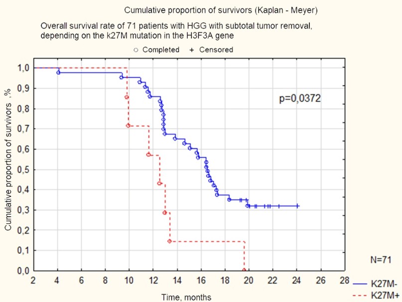
Figure 4: The survival rate of 71 patients with HGG who underwent subtotal tumor removal or biopsy, depending on the presence of the K27M mutation in the H3F3A gene.

No reliable prognostic significance of the above-mentioned mutations was revealed in the group of our HGG patients, who had a cystic tumor pattern. In other words, it was found that in the presence of cysts in the tumor according to MRI, the prognosis was poor regardless of the the mutation profile (p=0.2664) (Fig. 5).
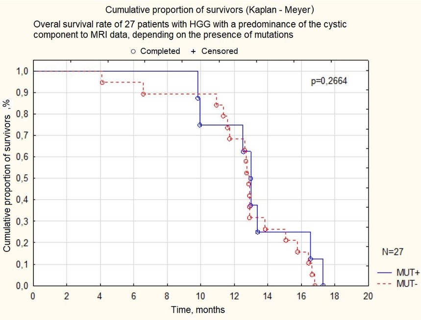
Figure 5: The overall survival rate of 27 patients with cystic HGG transformation according to initial MRI data, depending on the mutation status of the H3F3A, BRAF and IDH1 genes.

The modern molecular biological studies may have both a prognostic and diagnostic role. To date, HGG with the H3K27M mutation has a more aggressive clinical course in comparison with HGG with another genetic profile. Tumors with histone H3 K27M mutations are characterized by completely different clinical, radiological, and histological features. At the same time, the presence of BRAF and IDH-mutant HGG is predictor of better survival for the same group of tumors. In a comparative assessment of the survival of 18 children with HGG, depending on the presence or absence of mutations in the H3F3A, BRAF and IDH1 genes, it was found that patients with the K27M mutation in the H3F3A gene had significantly worse survival rates compared with patients with mutations in the BRAF and IDH1 genes (p= 0.0295) (fig. 6).
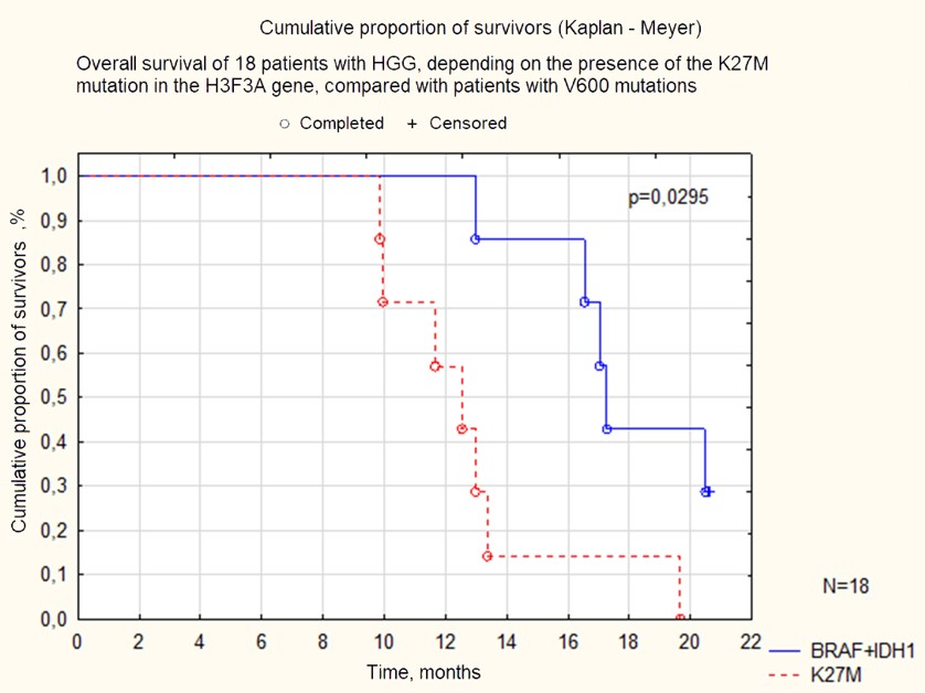
Figure 6: Overall survival of 18 patients with HGG, depending on the presence of the K27M mutation the H3F3A gene compared with patients with mutations in the BRAF and IDH1 genes.

Table 1: General characteristics of the analyzed group.

Discussion

HGG is usually a rapidly progressing disease that cannot be cured. Little progress has been made in treatment approaches over the past four decades, but the outcome of the disease remains disappointing: less than 20% of patients survive [21]. According to the generally accepted opinion, surgery, along with adjuvant radiation therapy and/or chemotherapy, plays a key role in the treatment of HGG, however, many treatment regimens, including monochemotherapy and combined chemotherapy, have not yet brought significant benefits in increase of OS in this group of patients. The goal of surgical intervention in HGG is to reduce the volume as safely as possible, this approach is associated with a better OS. Survival benefits have been reported in studies in which at least 70-80% of tumor tissue removal was achieved according to MR data [22-24]. In addition, total resection prevents false negative results caused by the limited number of tissue samples during biopsy and, thus, improves histological diagnosis to optimize adjuvant chemoradiotherapy [25]. Although complete total resection, defined as the radical removal of an MR contrast tumor, is associated with the remarkable increase in survival [26,27], it is often surgically difficult to achieve due to the infiltrative growth of this type of tumor. An increase in resection volume also leads to significant risks of postoperative neurological deficit. Surgical intervention is often difficult due to the extent of the lesion, localization, the presence of critical brain structures and the nature of tumor growth, which can lead to a deterioration of a patient condition. At the same time, even the presence of a histological diagnosis does not significantly affect the choice of treatment method and the prognosis of the disease in most cases of pediatric HGG.

Despite many modern methods of radiological examination, such MR signs as enhanced accumulation of contrast, limited diffusion, the presence of cysts and exophytic components remain controversial. There are still problems with the diagnostic algorithm of differential diagnosis. Therefore, it is so important to include clinical and anamnestic data along with radiological semiotics into the examination plan. This will help to make a correct diagnosis, nevertheless, extended diagnostic search is required in cases with an atypical picture or suspicion of a rare disease. An adequate approach to the examination of such patients requires a multidisciplinary team involving neurologists, radiologists, oncologists, neurosurgeons, and morphologists.

Over the past two decades, genomics has enriched and complemented traditional histopathology methodologies: DNA and RNA sequencing, RNA expression profiling, in situ fluorescence, hybridization, and, finally, DNA methylation research. These techniques have proven to be valuable tools for clarifying and improving both the classification and diagnosis of brain tumors in adults and children. The use of genomic and epigenomic molecular profiling methods has revealed the complex biological landscape of all forms of brain tumors, which has enriched knowledge in the field of pediatric neuro-oncology. We have moved from morphological to molecular categorization of diseases, in which we can now identify subgroups of tumors characterized by different clinical behavior, prognosis, anatomical localization and age of manifestation [28]. It seems that further accumulation of clinical material and expansion of the research on the molecular features of tumors is the most realistic way to improve HGG treatment methods.

HGG in children are not the same as the adult malignant gliomas [29]. The discovery of K27M mutation of the H3F3A gene significantly worsens survival rates and is associated with a more aggressive course of disease. Unfortunately, in our study the results of combined treatment were also unsatisfactory, and corresponded to global experience - H3K27 mutant HGG are always aggressive tumors for which there is no effective treatment other than palliative chemoradiotherapy aimed at increasing life expectancy.

Conclusions

  1. One of the most important prognostic features in brain gliomas is the grade of the tumor.
  2. When using complex modern methods of treatment, namely surgical treatment in combination with subsequent chemoradiotherapy in children with HGG, in the vast majority of cases it is possible to achieve OS in terms of up to 2 years.
  3. Patients with HGG who have a solid tumor structure on preoperative MRI of the brain and do not have the K27M mutation in the H3F3A gene have a better prognosis for life expectancy compared with the group of children with the presence of cysts on primary MRI and the K27M mutation in the H3F3A gene (p=0.000).
  4. The most unfavorable combination of prognostic factors in HGG (Grade III-IV) are: non-radical surgery, signs of tumor disintegration on the initial MRI scan, and the presence of a mutation in the K27M H3F3A gene.
  5. Considering both histological and molecular characteristics is important for accurate prognosis and optimal treatment plan for patients with CNS tumors, according to the WHO clinical recommendations 2021. We believe that the combination of the radiological features and the molecular genetic profile of the tumor has quite high prognostic significance, allowing, along with other factors, to personalize treatment approaches.

References

  1. Siegel DA; Richardson LC; Henley SJ; Wilson RJ; Dowling NF; Weir HK; et al. (2020). Pediatric cancer mortality and survival in the United States, 126(19):4379-4389.
  2. Nicholson JG; Fine HA. (2021). Diffuse Glioma Heterogeneity and Its Therapeutic Implications. Cancer Discov. 11(3):575-590.
  3. Jones C; Baker SJ. (2014). Unique genetic and epigenetic mechanisms driving paediatric diffuse high-grade glioma. Nat Rev Cancer. 14(10):10.1038/nrc3811.
  4. Louis DN; Perry A; Wesseling P; Brat DJ; Cree IA; Figarella-Branger D; et al. (2021). The 2021 WHO Classification of Tumors of the Central Nervous System: a summary. 23(8):1231-1251.
  5. Duffner PK; Horowitz ME; Krischer JP; Burger PC; Cohen ME; Sanford RA; et al. (1999). The treatment of malignant brain tumors in infants and very young children: an update of the Pediatric Oncology Group experience. Neuro Oncol. 1999 Apr;1(2):152-161.
  6. Davis ME. (2016).  Glioblastoma: Overview of Disease and Treatment. Clin J Oncol Nurs. 20(5 Suppl):S2-8.
  7. Zaitseva M.A; Shekhtman A.P; Papusha L.I; Valiakhmetova E.F; Yasko L.A; Druy A.E. (2020). Analysis of genetic aberrations in high-grade gliomas in children. Advances in Molecular Oncology. 7(3):37-47.
  8. Stupp R; Mason WP; van den Bent MJ; Weller M; Fisher B; Taphoorn MJ; et al. (2005). European Organisation for Research and Treatment of Cancer Brain Tumor and Radiotherapy Groups; National Cancer Institute of Canada Clinical Trials Group. Radiotherapy plus concomitant and adjuvant temozolomide for glioblastoma. 352(10):987-996.
  9. Castel D; Philippe C; Calmon R; Le Dret L; Truffaux N; Boddaert N; et al. (2015).  Histone H3F3A and HIST1H3B K27M mutations define two subgroups of diffuse intrinsic pontine gliomas with different prognosis and phenotypes. Acta Neuropathol. 130(6):815-827.
  10. Wang L; Li Z; Zhang M; Piao Y; Chen L; Liang H; et al. (2018).  H3 K27M-mutant diffuse midline gliomas in different anatomical locations. Hum Pathol. 78:89-96.
  11. Fanelli GN; Grassini D; Ortenzi V; Pasqualetti F; Montemurro N; Perrini P; et al. (2021).  Decipher the Glioblastoma Microenvironment: The First Milestone for New Groundbreaking Therapeutic Strategies. Genes (Basel). 12(3):445.
  12. Yao B; Delaidelli A; Vogel H; Sorensen PH. (2023). Pediatric Brain Tumours: Lessons from the Immune Microenvironment. 30(5):5024-5046.
  13. Ostrom QT; Gittleman H; Truitt G; Boscia A; Kruchko C; Barnholtz-Sloan JS. (2018). CBTRUS Statistical Report: Primary Brain and Other Central Nervous System Tumors Diagnosed in the United States in 2011-2015. Neuro Oncol. 20(suppl_4):iv1-iv86.
  14. Cerretti G; Cecchin D; Denaro L; Caccese M; Padovan M; Zagonel V; Lombardi G. (2023). Impressive response to dabrafenib and trametinib plus silybin in a heavily pretreated IDH wild-type glioblastoma patient with BRAFV600E -mutant and SOX2 amplification. Anticancer Drugs. 190-193. Epub 2022 Aug 10. PMID: 35946547.
  15. Gierke M; Sperveslage J; Schwab D; Beschorner R; Ebinger M; Schuhmann MU; Schittenhelm J. (2016). Analysis of IDH1-R132 mutation, BRAF V600 mutation and KIAA1549-BRAF fusion transcript status in central nervous system tumors supports pediatric tumor classification. J Cancer Res Clin Oncol. 89-100. Epub 2015 Jun 27. PMID: 26115961.
  16. Kadyrov Sh.U. (2022). Gliomas of deep brain structures. Specialty 3.1.10. Neurosurgery: Dissertation for the degree of Doctor of Medicine. Moscow, 2022. 277 p.
  17. Ryzhova M.V: Shishkina L.V. (2015). Use of molecular methods in diagnostics of poorly differentiated malignant brain tumors in children. Journal of Neurosurgery named after N.N. Burdenko. 79(2):10 20.
  18. Pollack IF; Hamilton RL; Sobol RW; Nikiforova MN; Lyons-Weiler MA; LaFramboise WA. (2011). Children's Oncology Group. IDH1 mutations are common in malignant gliomas arising in adolescents: a report from the Children's Oncology Group. 27(1):87-94. Epub 2010 Aug 20. PMID: 20725730; PMCID: PMC3014378.
  19. Toll SA; Tran HN; Cotter J; Judkins AR; Tamrazi B; Biegel JA; et al. (2019).  Sustained response of three pediatric BRAFV600E mutated high-grade gliomas to combined BRAF and MEK inhibitor therapy. Oncotarget. 551-557. PMID: 30728904; PMCID: PMC6355184.
  20. Clinical guidelines of the Russian Federation "Primary tumors of the central nervous system". Russian Federation. 2020. Available at: https://cr.minzdrav.gov.ru/recomend/578_1. Date of access: 17.03.2024
  21. Adamski J; Tabori U; Bouffet E. (2014).  Advances in the management of paediatric high-grade glioma. Curr Oncol Rep. 16(12):414. PMID: 25410415.
  22. D'Amico RS; Englander ZK; Canoll P; Bruce JN. (2017).  Extent of Resection in Glioma-A Review of the Cutting Edge. World Neurosurg. 103:538-549. Epub 2017 Apr 17. PMID: 28427971.
  23. Sanai N; Polley MY; McDermott MW; Parsa AT; Berger MS. (2011) . An extent of resection threshold for newly diagnosed glioblastomas. J Neurosurg. 2011 Jul;115(1):3-8.
  24. Chaichana KL; Jusue-Torres I; Navarro-Ramirez R; Raza SM; Pascual-Gallego M; Ibrahim A et al. (2014). Establishing percent resection and residual volume thresholds affecting survival and recurrence for patients with newly diagnosed intracranial glioblastoma. 113-122.
  25. Pang BC; Wan WH; Lee CK; Khu KJ; Ng WH. (2007). The role of surgery in high-grade glioma--is surgical resection justified? A review of the current knowledge. Ann Acad Med Singap. 36(5):358-363.
  26. Brown TJ; Brennan MC; Li M; Church EW; Brandmeir NJ; Rakszawski KL; et al. (2016). Association of the Extent of Resection With Survival in Glioblastoma: A Systematic Review and Meta-analysis. 2(11):1460-1469.
  27. Han Q; Liang H; Cheng P; Yang H; Zhao P. (2020). Gross Total vs. Subtotal Resection on Survival Outcomes in Elderly Patients With High-Grade Glioma: A Systematic Review and Meta-Analysis. 10:151.
  28. Clarke M; Mackay A; Ismer B; Pickles JC; Tatevossian RG; Newman S; et al. (2020). Infant High-Grade Gliomas Comprise Multiple Subgroups Characterized by Novel Targetable Gene Fusions and Favorable Outcomes. 10(7):942-963.
  29. McCrea HJ; Bander ED; Venn RA; Reiner AS; Iorgulescu JB; Puchi LA; et al. (2015). Sex, Age, Anatomic Location, and Extent of Resection Influence Outcomes in Children With High-grade Glioma. Neurosurgery.443-452; discussion 452-3.
TOP