
From Pain to Paralysis - Spontaneous Spinal Epidural Hematoma: A Case Report and Review of the Literature
Saqlain Anwar, MBBS1, Asim Ishfaq2, Ahmad Hassan, MBBS3, Aseef Ullah Chaudhary1, Hassan Waqar, MBBS1, Zohab Ahmed, MBBS4
¹House Officer, PNS Shifa Hospital Karachi, Pakistan
²Consultant Neurosurgeon, PNS Shifa Hospital Karachi
³Inter, Agha Khan University Hospital Karachi, Pakistan
⁴House Officer, Agha Khan University Hospital Karachi, Pakistan
*Corresponding author
Asim Ishfaq, Consultant Neurosurgeon, PNS Shifa Hospital Karachi
DOI: 10.55920/JCRMHS.2025.10.001449
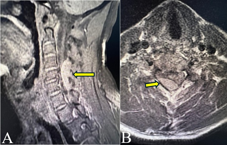
Figure 1: Preoperative MRI of cervical spine T2-weighted image with a hypertense signal in sagittal (A) and axial (B) cuts showing an extradural compression extending from the C4-C6 level as indicated by the yellow arrow.
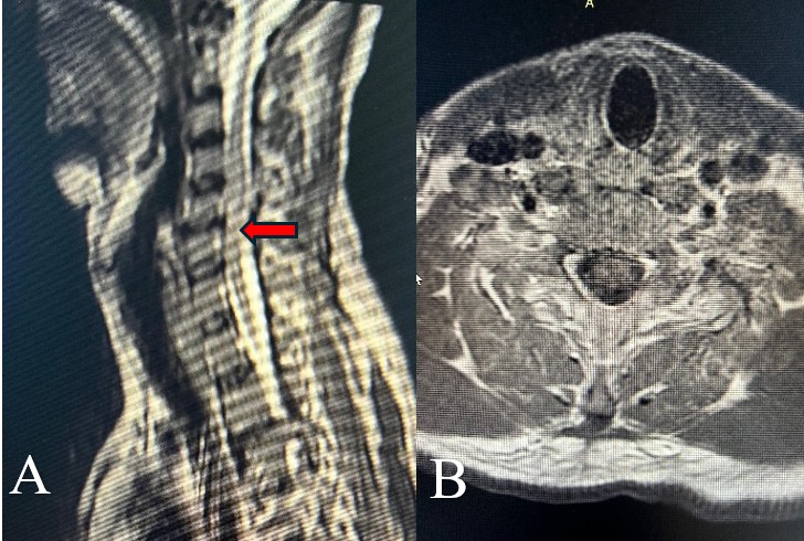
Figure 2: Postoperative sagittal (A) and axial (B) MRI images showing resolution of extradural hematoma compression with some residual edema at the C4-C6 level and a persistent T2- hyperintense signal in the spinal cord as indicated by the red arrow.
Given the severe spinal cord compression associated with impaired sensory and motor functions, the patient was planned for emergency C4-C6 laminectomy along with evacuation of the hematoma. A well-circumscribed hematoma was identified in the epidural space compressing the thecal sac and spinal cord. The hematoma was evacuated relieving the pressure on the spinal cord. There was no other pathology identified intraoperatively. After the surgery, the patient was shifted to the surgical ICU for observation. The patient was hypotensive for which he was given ionotropic support.
A postoperative MRI performed two days later showed the removal of the posterior elements of C4-C6 with the resolution of extradural hematoma compression. However, as shown in Figure 2, there was a persistent T2 hyperintense signal in the spinal cord. The patient's hypotension was stabilized, and he was transferred back to the surgical ward for ongoing management.
The patient showed partial improvement and after one week of hospital stay, he was discharged with recommendations for outpatient follow-up in one month. He was referred to his nearby hospital for physiotherapy and further monitoring. Upon discharge, his pain was significantly better, but his strength was partially recovered with a power of 3/5 in all limbs.
Timeline: The patient presented to PNS Shifa Hospital on 17-8-24 with complaints of cervicalgia and paraparesis. Contrast-enhanced MRI was performed the following day and C4-C6 decompressive laminectomy with the evacuation of the hematoma was done on 21-8-24. Two days later post-op CEMRI was done, and the patient was discharged after 1 week.