
De Garengeot Hernia: The Importance of Preoperative Diagnosis and Tailored Surgical Management
Yuechuan Liu† , Mingwei Gao† , Liming Wang*
Division of Hepatobiliary and Pancreatic Surgery, Department of General Surgery, The Second Affiliated Hospital of Dalian Medical University, Dalian, Liaoning, China.
†These authors have contributed equally to this work and share first authorship.
*Corresponding author
Liming Wang, Division of Hepatobiliary and Pancreatic Surgery, Department of General Surgery, The Second Affiliated Hospital of Dalian Medical University, Dalian, Liaoning, China.
DOI: 10.55920/JCRMHS.2025.10.001446
Abstract
De Garengeot hernia is an uncommon type of femoral hernia where the appendix is found within the hernial sac. The clinical presentation is often atypical, ranging from asymptomatic to severe pain due to incarceration or strangulation. We report a rare case of a middle-aged female patients who presented with a painful inguinal mass, which was initially suspected to be an incarcerated inguinal hernia. Preoperative CT imaging revealed a De Garengeot hernia with the appendix and mesoappendix within the hernial sac. The patient underwent a successful inguinal approach for hernia repair with appendectomy and mesh reinforcement. The use of preoperative imaging was crucial in guiding the surgical approach and ensuring a successful outcome. The operative strategy, challenges encountered, and recommendations for managing similar cases are discussed. With careful preoperative assessment and a tailored surgical approach, De Garengeot hernia can be managed effectively with favorable outcomes.
Keywords: De Garengeot Hernia; Inguinal Hernia; Femoral Hernia; Preoperative diagnosis; Surgical management
Background
De Garengeot hernia is a rare condition in which the appendix becomes entrapped within a femoral hernia sac1. The clinical presentation of this condition is often atypical, with nonspecific imaging findings that can make preoperative diagnosis challenging. Consequently, the diagnosis is frequently confirmed only during surgery. This report details a case of De Garengeot hernia in a 59-year-old female patient, accompanied by a thorough review of the relevant literature. The aim of this article is to provide a comprehensive analysis of the clinical presentation, diagnostic challenges, and treatment strategies associated with this rare hernia, offering valuable guidance for clinicians who may encounter similar cases in their practice.
Case Presentation
A 59-year-old female presented with a one-day history of persistent pain and enlargement of a previously reducible right inguinal mass, first noted 12 years ago. She had a history of right adnexectomy for an ovarian cyst. Physical examination revealed a 5 cm × 3 cm mass in the right inguinal region. Laboratory tests showed a white blood cell count of 9.34×10⁹/L (reference range: 3.5-9.5×10⁹/L). Ultrasound suggested a cystic mass, and CT confirmed a right inguinal hernia with suspected bowel loops as contents (Figure 1). She was taken to surgery for suspected incarcerated inguinal hernia. A right inguinal oblique incision revealed a 4 cm × 5 cm hernia sac containing the appendix and mesoappendix, with ischemic necrosis of the mesoappendix and blood-stained fluid.
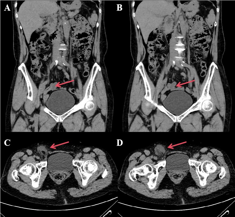
Figure 1: Preoperative CT images (A, B) Preoperative CT images showing appendix reconstruction. (C, D) Preoperative thin-slice CT scans
The hernia sac protruded from the femoral canal, confirming a femoral hernia. An appendectomy and partial hernia sac resection were performed. As there was no purulent perforation or bowel obstruction, the defect was repaired using a modified Kugel patch. The patient had an uneventful recovery and was discharged on the fourth postoperative day.
Discussion
Recent advancements in CT technology, including thin-slice imaging and appendix reconstruction, along with increased awareness of De Garengeot hernia, have significantly improved diagnostic accuracy. Since Takemura et al. first used CT for its preoperative diagnosis in 2000, the reported sensitivity of CT in diagnosing De Garengeot hernia is approximately 61%2,3 Three key CT findings are important: a tubular structure ventral and medial to the femoral vein, continuity with the cecum, and a blind-ended structure4. Despite this, a definitive diagnosis may still only be confirmed intraoperatively. In our case, preoperative CT confirmed two of these criteria and revealed gas within the hernia sac, raising suspicion of bowel loop incarceration. This led us to suspect De Garengeot hernia, though we did not rule out small bowel obstruction. Surgery confirmed our diagnosis without evidence of bowel obstruction.
The surgical management of De Garengeot hernia requires an individualized approach, guided by the patient’s clinical presentation, imaging findings, and potential complications such as ischemia or infection. The preferred initial method is typically a single inguinal incision, which allows for both effective hernia repair and safe appendectomy, particularly when the appendix is uncomplicated and easily accessible5-7.
In more complex cases, such as those involving bowel obstruction or abscess formation, an open laparotomy may be necessary to ensure full exposure and complete reduction of the hernia8. The choice of incision—McBurney or midline—should be based on anatomical and pathological findings.
Laparoscopic surgery offers a valuable alternative, especially for clinically stable patients with a clear preoperative diagnosis. It enables a thorough examination of the abdominal cavity and can reduce postoperative pain, speed recovery, and lower the risk of wound complications9,10. However, laparoscopic repair must be approached cautiously in the presence of significant inflammation, gangrene, or perforation, as these factors can complicate the procedure.
In some cases, combining an inguinal approach with laparoscopy provides greater flexibility, particularly when the appendix cannot be adequately accessed through the inguinal route alone11.
The decision to use mesh in hernia repair is critical. When the surgical field is uncontaminated, mesh repair is preferred due to its lower recurrence risk12,13. In cases of severe inflammation or infection, tissue-based repair may be more appropriate to mitigate the risk of mesh-related complications. Surgeons should also consider protective measures such as drain placement or delayed wound closure, particularly in cases involving necrosis or perforation14. In this case, preoperative imaging strongly suggested De Garengeot hernia, though the presence of purulent perforation remained unclear. Intraoperatively, sterile ischemic necrosis of the mesoappendix was confirmed, allowing safe use of mesh following resection of the hernia sac and appendix, leading to an optimal outcome.
The successful management of De Garengeot hernia requires a thorough understanding of its atypical presentation and the potential complexities involved in its diagnosis and treatment. This case underscores the importance of integrating detailed preoperative imaging with careful intraoperative decision-making to tailor the surgical approach to the patient’s specific needs. The use of mesh in hernia repair, when appropriate, can significantly reduce the risk of recurrence and support better patient outcomes. However, in the presence of complicating factors such as ischemia or infection, a more cautious approach is warranted.
While this case report includes comprehensive preoperative CT images that were instrumental in guiding the surgical strategy, it lacks intraoperative images. This limitation is due to the urgency of the clinical situation, where the primary focus was on providing timely and effective surgical care rather than on documentation. Despite this, the preoperative imaging provided clear evidence that strongly supported the diagnosis and informed the surgical approach, ultimately leading to a successful outcome.
Conclusion
De Garengeot hernia is a rare condition with atypical presentation. Accurate preoperative diagnosis is crucial for guiding surgical decision-making and ensuring successful outcomes. Detailed CT imaging allows for a tailored approach, including the safe use of mesh, leading to an effective repair. This highlights the importance of thorough preoperative assessment in managing complex hernias efficiently.
Author contributions: Liming Wang: Concepts, design, definition of intellectual content, clinical studies, manuscript review; Yuechuan Liu: Literature search, clinical studies, data acquisition, manuscript preparation, manuscript review; Mingwei Gao: Manuscript editing, manuscript review, data analysis, manuscript preparation.
Ethics approval and consent to participate: The studies involving human participants underwent a thorough review and received approval from the ethics committee of The Second Hospital of Dalian Medical University.
Data availability statement: The data supporting the findings of this study are accessible from the corresponding author upon a reasonable request.
Financial support and sponsorship: Nil.
Conflicts of interest: The authors declare that they have no competing interests.
References
- de Garengeot RJC. Traite des operations de chirurgie. vol 1. Chez Huart; 1731.
- Takemura M, Iwamoto K, Goshi S, Osugi H, Kinoshita H (2000). Strangulated femoral hernia containing gangrenous appendicitis: report of a case; 25(5):789-791.
- Guenther TM, Theodorou CM, Grace NL, Rinderknecht TN, Wiedeman JE (2021). De Garengeot hernia: a systematic review. Surg Endosc. Feb; 35(2):503-513. doi:10.1007/s00464-020-07934-5
- Tsuruta S, Miyake H, Nagai H, Yoshioka Y, Yuasa N, Fujino M (2021). Clinicopathological characteristics of De Garengeot hernia: six case reports and literature review. Surg Case Rep. Jan 11; 7(1):14. doi:10.1186/s40792-020-01098-z
- Couto HS, de Figueiredo LO, Meira RC, et al (2016). Treatment of De Garengeot’s hernia using De Oliveira’s technic: A case report and review of literature. International Journal of Surgery Case Reports; 26:57-60.
- Jin Z, Imtiaz MR, Nnajiuba H, Samlalsingh S, Ojo A (2016). De Garengeot's Hernia: Two Case Reports with Correct Preoperative Identification of the Vermiform Appendix in the Hernia. Case Rep Surg: 2424657.
- Nnadozie UU, Onyeyirichi O, Maduba CC, Ekwesianya AC (2021). Obstructed right Duari hernia. Ghana medical journal. Sep; 55(3):229-231. doi:10.4314/gmj.v55i3.9
- Julien B, Huang Y, Ooi WL, Beck M (2022). Management of appendicitis in a De Garengeot hernia: Lockwood approach. BMJ Case Rep. Jul 6; 15(7)
- Beysens M, Haeck L, Vindevoghel K (2013). Laparoscopic appendectomy combined with TEP for de Garengeot hernia: case report. Acta Chir Belg. Nov-Dec; 113(6):468-70.
- Leite TF, Chagas CA, Pires LA, Cisne R, Babinski MA (2016). De Garengeot's hernia in an 82-year-old man: a case report and clinical significance. J Surg Case Rep. Jul 5; 2016(7)
- Linder S, Linder G, Mansson C (2019). Treatment of de Garengeot's hernia: a meta-analysis. Hernia. Feb; 23(1): 131-141.
- Kokoszka M, Wojtowicz J (2015). De Garengeot's Hernia; Acute Appendicitis In An Incarcerated Femoral Hernia. Pol Przegl Chir. Jul 1; 87(7): 365-7.
- Yu PC, Wang LT, Chang CY, Tsai YC, Chong KH (2024). De Garengeot Hernia, an acute appendicitis in the right femoral hernia canal, and successful management with transabdominal closure and appendectomy: a case Report. BMC urology. Feb 16; 24(1): 41.
- Vos CG, Mollema R, Richir MC (2017). De Garengeot hernia: an uncommon presentation of acute appendicitis. Acta Chir Belg. Feb; 117(1):52-54.