
Microsurgery and Coil Occlusion Tackle Fistulas
Erekle Ekvtimishvili1, Arpita Meher2*, Jessen Johnson2
1MD, PhD, Assistant Professor at TSMU, Head of Endovascular Neurosurgery Department, High Technology Medical Center, University Clinic, Tbilisi, Georgia
2Department of Medicine, Tbilisi State Medical University, Tbilisi, Georgia
*Corresponding author
*Arpita Meher, Department of Medicine, Tbilisi State Medical University, Tbilisi, Georgia
DOI: 10.55920/JCRMHS.2025.10.001415
Abstract
This revolutionary study report presents a convincing example of a 47-year-old guy who developed Carotid-Cavernous Fistula (CCF) as a result of a traffic accident. Our approach comprised ligating the internal carotid artery and inserting a transarterial coil, which was a novel mix of microsurgical and endovascular techniques. The patient's pulsatile exophthalmos and ocular bruit resolved quickly, indicating that the fistula had been successfully repaired. This research delves into CCF classifications, etiology, and pathophysiology, shedding insight into the complex diagnostic process of color Doppler imaging and angiography. Treatment alternatives, focusing on endovascular procedures, are explored, offering a thorough understanding of the changing landscape of CCF therapy. This work is a huge step forward in enhancing therapy options for CCFs, demonstrating the dynamic interaction between microsurgery and cutting-edge endovascular interventions.
Keywords: Carotid-Cavernous Fistulas (CCFs), Chemosis, Digital Subtraction Angiography (DSA), Arteriovenous Fistula
Abbreviations: Carotid-Cavernous Fistula (CCF), Computed Tomography Angiography (CTA), Magnetic Resonance Angiography (MRA), Internal Carotid Artery (ICA), Inferior Petrosal Sinus (IPS), Persistent Primitive Trigeminal Artery (PPTA), Intraocular Pressure (IOP)
Introduction
Carotid Cavernous Fistulas (CCFs) are classified according to their origin and flow characteristics. Whether spontaneous or trauma-induced, they frequently manifest with specific clinical characteristics such as pulsatile exophthalmos and ocular bruit. Imaging methods diagnose the fistula; therapy is determined by its nature and severity. This case demonstrates the effective combination of microsurgical and endovascular procedures, offering important insights into the management of CCF.
This case report describes the presentation, diagnosis, and effective treatment of a 47-year-old Caucasian male. He was diagnosed with CCF with CTA. The treatment technique used a combination of microsurgical and endovascular treatments. The treatment resulted in the closure of the fistula and the patient's spontaneous recovery.
Case Presentation
A 47 y/o Caucasian male was presented to the ER at High Technology Medical Center University Clinic, following a road traffic accident, with a severe headache and throbbing eye pain, occasionally affecting his visual clarity.
Upon thorough examination, the patient exhibited chemosis, pulsatile exophthalmos, and an ocular bruit, collectively constituting the classical symptomatic triad indicative of Carotid-Cavernous Fistulas (CCFs). Based on Barrow classification it is Type A.
Subsequent cerebral angiography confirmed the presence of a CCF originating from the Right Internal Carotid Artery, accompanied by multiple traumatic injuries in the Cavernous Sinus. In the conventional DSA image [Fig.1], the Left Internal Carotid Artery (marked with a blue arrow) is visible, while the Carotid-Cavernous Fistula (marked with a red arrow) involving the Right Internal Carotid Artery and the Cavernous sinus is also identifiable. In the second DSA view [Fig.2], the Carotid-Cavernous Fistula (CCF) is observed to drain into both the Superior and Inferior Ophthalmic veins, indicating complex vascular involvement and potential clinical implications.
In the conventional DSA below [Fig.3] of the vertebral basilar system, the presence of the CCF is evident, highlighting the need for detailed assessment and management of vascular abnormalities in this region.
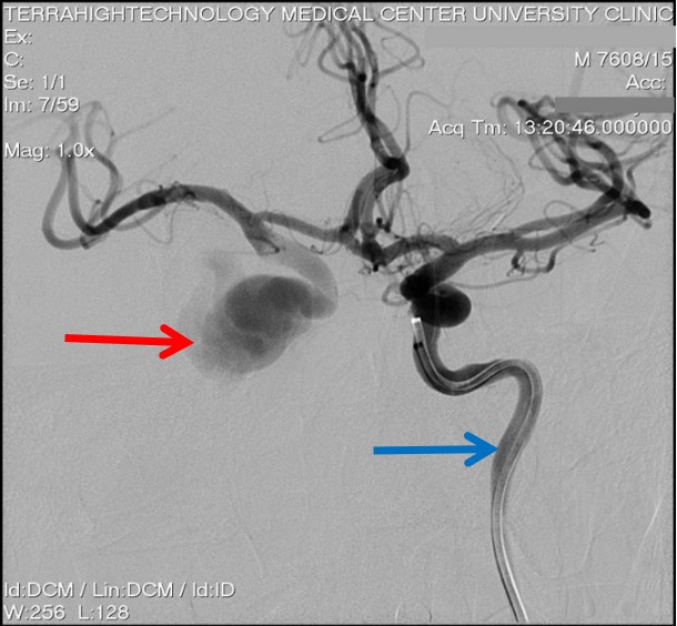
Figure 1: View of conventional DSA of the Left Internal Carotid Artery (blue arrow). The Carotid-Cavernous Fistula (red arrow) of the Right Internal carotid artery and the Cavernous sinus can be identified.
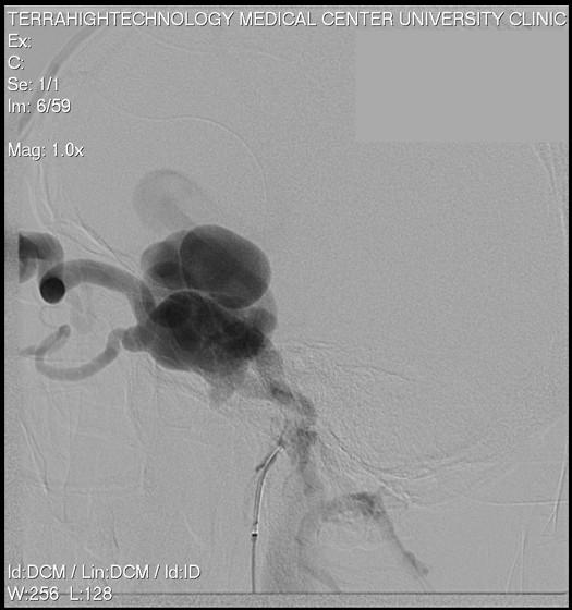
Figure 2: View of DSA showing the CCF draining into the Superior and Inferior Ophthalmic Vein.
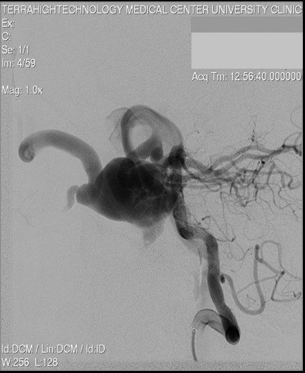
Figure 3: View of conventional DSA of the vertebral basilar system indicating the CCF.
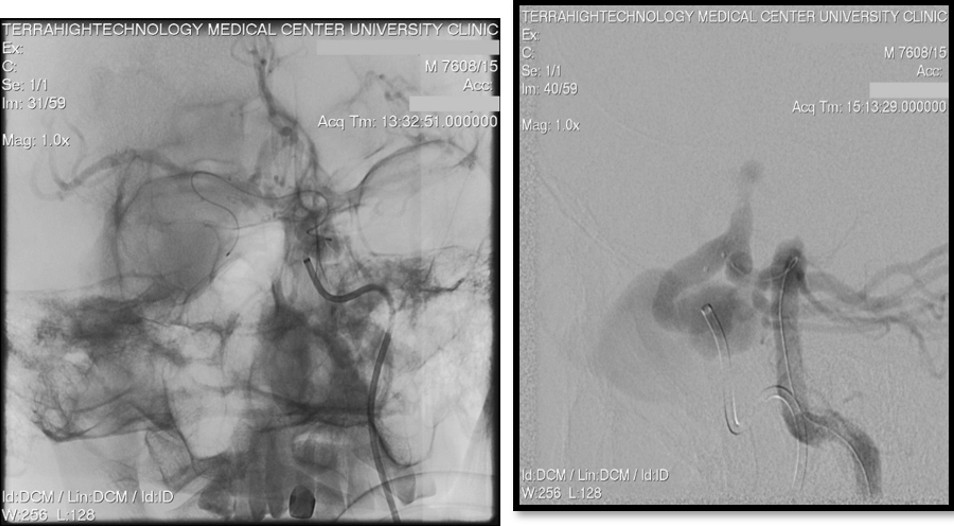
Figure 4: Ligation of the Right internal carotid artery and the microcatheterization through the Anterior Communicating and Posterior Communicating to introduce coils into the fistula
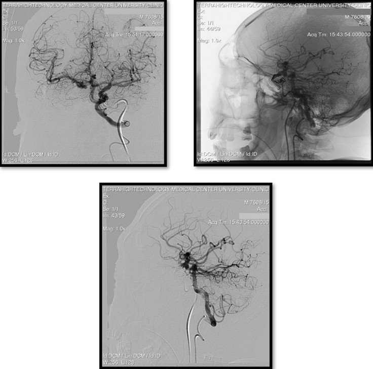
Figure 5: After transarterial coil embolization, the total filling of the Right Hemisphere can be noted from the Left Internal Carotid artery and the vertebral basilar system
It was treated using a combination of microsurgical and endovascular procedures. The microsurgical element included ligating the ICA using a standard surgical approach, which interrupted the aberrant blood flow caused by the fistula. Similarly, the endovascular component used a transarterial technique, with dual microcatheterization into the anterior communicating artery and posterior communicating artery. During this endovascular phase, coils were carefully inserted to occlude and block the aberrant arteriovenous link, delivering a comprehensive and successful therapy for Carotid Cavernous Fistula. Resulting in the closure of the fistula and the patient's spontaneous recovery. The procedure involved ligation of the Right internal carotid artery and microcatheterization through both the Anterior Communicating and Posterior Communicating arteries to introduce coils into the fistula, aiming to effectively occlude the abnormal vascular connection and restore normal blood flow dynamics as demonstrated in [Fig.4].
Following the procedure, the patient's pulsatile exophthalmos and ocular bruit rapidly resolved within a few hours, while subsequent follow-up revealed a gradual but consistent improvement in the patient's chemosis. The images below [Fig.5] shows transarterial coil embolization, comprehensive filling of the Right Hemisphere is observed, facilitated by blood flow redirection from both the Left Internal Carotid artery and the vertebral basilar system, indicating successful closure of the fistula and restoration of cerebral perfusion dynamics.
Discussion
Arteriovenous abnormalities between the carotid artery or its branches and the cavernous sinus are known as carotid-cavernous fistulas (CCFs). Direct or indirect, high or low flow, or Barrow types A, B, C, or D are typical categories [1]. Trauma or spontaneous development are two possible causes of these lesions [2].
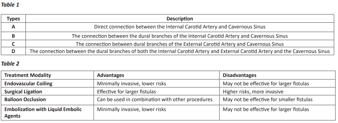
Classification: The two types of CCFs are direct fistulas (Barrow type A) and dural or indirect fistulas (Barrow types B, C, and D).
Types of Carotid-Cavernous Fistulas and Their Description [3]
Indirect fistulas often have low flow, while direct fistulas typically have high flow. The most prevalent kind of spontaneous dural carotid-cavernous fistulas is type D. The artery of the inferior cavernous sinus is the most commonly involved trunk of the Internal Carotid Artery (ICA), however, dural fistulas can also involve the meningohypophyseal trunk and its branches. The internal maxillary artery is the most usually involved branch of the external carotid artery, with additional implicated branches including the middle and accessory meningeal arteries, ascending pharyngeal artery, anterior deep temporal artery, and posterior auricular artery [3].
Aetiology: Several reasons can lead to direct fistulas, which are defined by abnormal connections between veins and arteries. These include forceful or penetrating trauma, such as concussions, that can damage normal vascular structures. Direct fistulas can also develop as a result of the rupture of an intracranial aneurysm within the cavernous sinus, a sophisticated network of veins near the base of the brain. A few genetic disorders, such as connective tissue disease Ehlers-Danlos syndrome type IV, make people more likely to develop direct fistulas. Furthermore, aberrant arteriovenous connections can be unintentionally formed by iatrogenic interventions such as internal carotid endarterectomy, transarterial endovascular procedures, and surgeries like craniofacial or trans-sphenoidal excision of pituitary tumors [3].
On the other hand, indirect or dural fistulas frequently have distinct underlying causes. Among these is hypertension, a disorder marked by elevated blood pressure, which may exacerbate blood vessel weakening and aberrant dilating, which may result in the development of fistulas. Dural fistulas have also been linked to Internal Carotid Artery (ICA) dissection, a condition in which there is a rupture in the inner lining of the carotid artery, and fibromuscular dysplasia, a disorder that affects the walls of arteries. Interestingly, direct and indirect fistulas are linked to Ehlers-Danlos syndrome type IV, which is characterized by abnormalities in connective tissue. Postmenopausal women are especially prone to these diseases, perhaps because of age-related vascular alterations and hormonal changes that might make them more vulnerable to the development of fistulas [3].
Pathophysiology: The thrombosis of cavernous sinus venous outflow channels, which is followed by vascular alterations to supply collateral flow, is most likely the initial cause of the pathophysiology of CCFs. Since it explains the formation of arteriovenous fistulas affecting different dural sinuses, this pathogenesis theory is widely accepted. Nonetheless, other authors contend that the dilating of pre-existing dural-arterial anastomoses results in the formation of CCFs following the rupture of one or more thin-walled dural arteries. The collateral blood supply that these anastomoses subsequently provide causes an angiographic result that is similar to a congenital vascular abnormality [3].
Symptoms: Carotid-cavernous fistulas (CCFs) are characterized by a variety of symptoms that are indicative of an irregular connection between the carotid artery and the cavernous sinus, a region that houses vital blood arteries and nerves. Subjective bruits, which are abnormal sounds produced inside the skull by turbulent blood flow, are frequently reported by patients. The changed vascular dynamics that affect the nerves responsible for eye movement can cause diplopia or double vision. Patients may also have redness in the eyes and weeping or tearing of the eyes, which is a sign of ocular congestion brought on by irregular blood flow. The increased pressure on ocular tissues might result in symptoms such as visual impairment and feelings of an ocular foreign body. Perhaps as a result of the vascular alterations and related pressure consequences, headaches are frequently experienced [3].
Ocular symptoms are more prevalent with anterior draining fistulas. Patients suffering from posteriorly draining fistulas may experience neurologic symptoms such as disorientation and expressive aphasia, as well as diplopia from isolated ocular motor nerve pareses. Clinical symptoms and signs are frequently intense in direct fistula and more indolent in cases of dural fistula [3].
The clinical manifestations of CCFs differ based on the lesion's flow dynamics, specifically on high or low flow. Proptosis, an abnormal protrusion of the eyeball, is one typical sign. In high-flow lesions, proptosis may pulse because of turbulent and fast blood flow through the abnormal connection between the carotid artery and the cavernous sinus. The conjunctival and episcleral arteries show arterialization, suggesting a change in blood flow dynamics, and the affected eye frequently appears red. Chemosis, or conjunctival swelling, is a common observation. Misalignment of the eyes, or strabismus, can be caused by orbital congestion, ocular motor nerve dysfunction, or a combination of the two. Upon inspection, an ocular bruit—an irregular sound brought on by turbulent blood flow—might be audible. Stasis retinopathy and elevated intraocular pressure (IOP) are symptoms of vascular congestion in the eye. In severe cases, a noticeably raised episcleral venous pressure may lead to the development of central retinal vein blockage. Due to the aberrant vascular dynamics linked to carotid-cavernous fistulas (CCF), acute trauma or ischemia may cause optic neuropathy, which can present as glaucomatous or non-glaucomatous. When taken as a whole, these clinical indicators emphasize the intricate neurological and ocular symptoms of CCF, stressing the significance of early identification and treatment [2,3]
Diagnosis: If a CCF is suspected, the assessment may involve color Doppler imaging, routine tonometry, pneumotonometry, and ultrasonography. Color Doppler detects flow direction and velocity in orbital veins to determine arterial flow in cases with CCF. For individuals in whom a carotid-cavernous fistula is suspected, neuroimaging is required. This may use non-invasive CTA or MRA. Whether the carotid-cavernous fistulas are diffuse or direct, both approaches are extremely sensitive to them when they cause visual problems. For individuals for whom a carotid-cavernous fistula is suspected, DSA may still be required. Digital subtraction angiography is still the gold standard for carotid-cavernous fistula classification and diagnosis, and it can be utilized for both diagnostic and therapeutic purposes.
The drainage pattern of the fistula is also described by Digital subtraction angiography, including whether it drains posteriorly via the inferior petrosal sinus (IPS), anteriorly via the superior ophthalmic vein, or both. Additionally, it can detect cerebral vein reflux [3].
Clinical Implications: The case describes a 55-year-old female of Chinese origin presenting with symptoms consistent with a carotid-cavernous fistula (CCF) secondary to a ruptured persistent primitive trigeminal artery (PPTA) aneurysm. The patient underwent successful endovascular embolization with detachable coils and an Onyx-18 liquid embolic system via a transarterial approach. This procedure effectively obliterated the fistula and aneurysm, leading to the resolution of symptoms and the absence of neurological deficits during hospitalization and follow-up. The patient experienced immediate relief from symptoms such as orbital bruit and chemosis post-embolization, with mild residual abducens nerve paresis resolving within a month. Endovascular embolization proved to be a safe and effective treatment modality for this complex vascular pathology, providing durable symptom relief and preventing recurrence without significant neurological sequelae [4]
Carotid-cavernous fistula (CCF) can present with atypical symptoms such as cerebral infarction without the typical ocular manifestations. Two cases presented here illustrate this phenomenon. In the first case, a patient with ICA stenosis developed a massive cerebral infarction due to an interruption of the ICA blood supply secondary to CCF. The second case involved a traumatic eye rupture resulting in CCF and subsequent cerebral infarction. Digital subtraction angiography (DSA) remains the gold standard for diagnosing CCF. While some patients with mild symptoms may benefit from ICA compression, the majority require endovascular treatment to embolize the fistula. Various embolic materials such as detachable balloons, laminated stents, spring rings, and Onyx can be used (5).
The treatment modality employed in these cases was endovascular embolization. In the first case, due to concerns about ischemia-reperfusion injury, the patient was managed conservatively with medications and rehabilitation therapy. In the second case, endovascular embolization was performed successfully to completely occlude the fistula. These cases underscore the importance of considering CCF in patients with head trauma and atypical symptoms of cerebral ischemia, with DSA being crucial for diagnosis and guiding appropriate treatment [5].
The patient presented with a gunshot wound to the posterior pharynx resulting in complex midface fractures, cervical spine injury, and traumatic CCF. Initial imaging with CT angiography revealed vascular trauma and subsequent DSA confirmed the presence of a carotid-cavernous fistula with rapid arteriovenous shunting. Due to unsuccessful attempts at endovascular access, the decision was made to ligate the internal carotid artery with endovascular coiling, resulting in the successful resolution of the fistula and continued perfusion via collateralization. The treatment modality, in this case, involved endovascular intervention with coil occlusion of the left internal carotid artery (ICA) to address the traumatic CCF. This approach aimed to halt the abnormal arteriovenous shunting and restore normal cerebral perfusion. Despite initial challenges in accessing the fistula, ligation of the ICA proved successful in resolving the carotid-cavernous fistula and preserving cerebral blood flow via collateralization from the contralateral circulation [6].
These examples show the variety of causes and manifestations of CCF, from vascular malformations such as persistent primitive trigeminal artery (PPTA) aneurysms to traumatic injuries. DSA is still essential for proper diagnosis even with differences in presentation. As the main therapeutic option, endovascular embolization is found to be effective in closing the fistula and preventing neurological consequences. Nonetheless, difficulties in gaining access to the fistula can need other methods, including internal carotid artery ligation, underscoring the significance of a customized therapeutic strategy. All things considered, these instances demonstrate how important it is to act quickly and use a multidisciplinary strategy to get the best possible results and avoid long-term issues related to CCF.
Treatment: The type of treatment is determined by the kind of CCF under investigation, clinical history, and other factors. The majority of low-flow or Barrow type B, C, or D arteriovenous fistulas cure on their own and don't require treatment; other approaches may require ongoing observation. A patient's symptom profile advances with low-flow CCFs. Due to the relatively high chance that the patient's symptom profile would worsen and result in a brain hemorrhage, all high-flow or Barrow type A fistulas should be treated [1].
The only options for managing direct CCFs were to either leave the fistula untreated by ligating the cervical ICA proximal to the fistula and the intracranial ICA distal to it or to observe the condition or occlude the common carotid artery or intracranial artery, both of which could result in a cerebral ischemic event due to an embolic event. With the development of endovascular interventional procedures, open surgical therapies are no longer the preferred approach. Endovascular treatment offers a lower risk of cerebral infarction and is less invasive than ICA sacrifice. The arterial supply, venous drainage, blood flow rate via the fistula, and Willis circle patency all influence the optimal treatment plan. In situations where there are significant ICA wall tears, which could allow the injected embolic material to return to the arterial circulation and put the patient at risk for embolic consequences, flow-diverting stent support may be used for endoluminal repair. To stop the backflow of the injected substance, these stents may be placed across the ICA tear [3].
Treatment options for dural CCFs include observation, IOP-lowering drugs, and stereotactic radiosurgery, intermittent compression of the ipsilateral internal carotid artery or superior ophthalmic vein, and endovascular intervention. If surgical surgery is not required, patients may utilize occlusion methods, such as external manual carotid compression, to facilitate the resolution of the CCF. While a careful waiting approach makes sense for many individuals with a dural CCF, treatment may occasionally be required to prevent long-term effects. Intervention is indicated by uncontrollably high intraocular pressure, persistent diplopia, and severe proptosis with corneal exposure, optic neuropathy, retinal ischemia, severe bruit, and cerebral venous drainage from the fistula. Endovascular therapy, which can be delivered intravenously or transarterially, is the first line of treatment [3].
Comparison of Treatment Modalities: A comprehensive overview of the various treatment modalities for carotid-cavernous fistula [7-9].
The importance of the findings of this clinical case are as follows:
- The case highlights the effectiveness of a multidisciplinary treatment approach combining microsurgical and endovascular techniques.
- The rapid resolution of symptoms such as pulsatile exophthalmos and ocular bruit, along with gradual improvement in chemosis, underscores the importance of regular clinical and radiological surveillance to ensure optimal patient outcomes.
- Successful closure of the CCF resulted in the restoration of normal cerebral perfusion dynamics, as evidenced by comprehensive filling of the right hemisphere on imaging studies.
Conclusion
In a compelling conclusion, this case demonstrates a victorious mix of artistry and inventiveness in treating CCF. Witnessing the miraculous recovery of a 47-year-old guy following a road traffic accident, our combined microsurgical and endovascular technique not only closed the fistula but also arranged a rapid symphony of relief from pulsatile exophthalmos and ocular bruit. Our trip through the complex labyrinth of CCF classifications, etiology, and pathophysiology carried us into the dynamic landscape of diagnostic procedures, where color Doppler imaging and angiography provided vivid strokes of clarity. As the focus switched to therapy, the developing dance underscored the importance of endovascular procedures, in sync with the throbbing rhythm of development.
Acknowledgments: None
Consent: Written informed consent was obtained from the patient for the publication of this case report and accompanying images.
Conflict of interest: No apparent conflicts of interest.
Ethical approval: The study is exempt from ethical approval in our institution.
Sources of funding: None available.
References
- Henderson A; Miller N (2018). Carotid-cavernous fistula: current concepts in aetiology; investigation; and management. Eye32; 164–172.
- Hasan S; Pedro AS &Mauricio JA (2021). Carotid Cavernous Fistula Treatment via Flow Diversion: A Systematic Review of the Literature; World Neurosurgery; Volume 149; Pages e369-e377; ISSN 1878-8750
- Pavlos T; Andreas T & Dimitrios X (2021). Carotid cavernous fistula (CCF) treatment approaches: A systematic literature review and meta-analysis of transarterial and transvenous embolization for direct and indirect CCFs; Clinical Neurology and Neurosurgery; Volume 204; 106601; ISSN 0303 8467.
- Henderson AD; Miller NR (2018). Carotid-cavernous fistula: current concepts in aetiology; investigation; and management. Eye (London; England); 32(2); 164–172.
- Sun P; Chai Y & Fang W (2022). Case report: Spontaneous carotid-cavernous fistula associated with persistent primitive trigeminal artery aneurysm rupture. Frontiers in neurology; 13; 923186.
- Jiang H; Zeng Q & Jiang W (2023). Carotid-cavernous sinus fistula with primary clinical manifestation of cerebral infarction: Description of two cases.
- Hamedani H; Hellmann D & Boyce W (2022). Traumatic carotid-cavernous fistula: A case report. Radiology case reports; 17(6); 1955–1958.
- Luis NGC; Rene AC &Alyssa A Botelho; Suzanne K. et al. Carotid-cavernous fistula | stroke.
- Al-Boqami B A; Tammar RS & Alharbi SE (2023). Endovascular Intervention for a Carotid-Cavernous Fistula: A Case Report. Cureus; 15(9); e44902.
- Sharma R; Ponder C & Kamran M (2022). Bilateral Carotid-Cavernous Fistula: A Diagnostic and Therapeutic Challenge. Journal of investigative medicine high impact case reports; 10; 23247096221094181.